EP3398554B1 - Prosthesis for inguinal hernia repair - Google Patents
Prosthesis for inguinal hernia repair Download PDFInfo
- Publication number
- EP3398554B1 EP3398554B1 EP17305489.1A EP17305489A EP3398554B1 EP 3398554 B1 EP3398554 B1 EP 3398554B1 EP 17305489 A EP17305489 A EP 17305489A EP 3398554 B1 EP3398554 B1 EP 3398554B1
- Authority
- EP
- European Patent Office
- Prior art keywords
- prosthesis
- medial
- inferior
- piece
- edge
- Prior art date
- Legal status (The legal status is an assumption and is not a legal conclusion. Google has not performed a legal analysis and makes no representation as to the accuracy of the status listed.)
- Active
Links
Images

Classifications
-
- A—HUMAN NECESSITIES
- A61—MEDICAL OR VETERINARY SCIENCE; HYGIENE
- A61F—FILTERS IMPLANTABLE INTO BLOOD VESSELS; PROSTHESES; DEVICES PROVIDING PATENCY TO, OR PREVENTING COLLAPSING OF, TUBULAR STRUCTURES OF THE BODY, e.g. STENTS; ORTHOPAEDIC, NURSING OR CONTRACEPTIVE DEVICES; FOMENTATION; TREATMENT OR PROTECTION OF EYES OR EARS; BANDAGES, DRESSINGS OR ABSORBENT PADS; FIRST-AID KITS
- A61F2/00—Filters implantable into blood vessels; Prostheses, i.e. artificial substitutes or replacements for parts of the body; Appliances for connecting them with the body; Devices providing patency to, or preventing collapsing of, tubular structures of the body, e.g. stents
- A61F2/0063—Implantable repair or support meshes, e.g. hernia meshes
- A61F2002/0068—Implantable repair or support meshes, e.g. hernia meshes having a special mesh pattern
-
- A—HUMAN NECESSITIES
- A61—MEDICAL OR VETERINARY SCIENCE; HYGIENE
- A61F—FILTERS IMPLANTABLE INTO BLOOD VESSELS; PROSTHESES; DEVICES PROVIDING PATENCY TO, OR PREVENTING COLLAPSING OF, TUBULAR STRUCTURES OF THE BODY, e.g. STENTS; ORTHOPAEDIC, NURSING OR CONTRACEPTIVE DEVICES; FOMENTATION; TREATMENT OR PROTECTION OF EYES OR EARS; BANDAGES, DRESSINGS OR ABSORBENT PADS; FIRST-AID KITS
- A61F2210/00—Particular material properties of prostheses classified in groups A61F2/00 - A61F2/26 or A61F2/82 or A61F9/00 or A61F11/00 or subgroups thereof
- A61F2210/0004—Particular material properties of prostheses classified in groups A61F2/00 - A61F2/26 or A61F2/82 or A61F9/00 or A61F11/00 or subgroups thereof bioabsorbable
-
- A—HUMAN NECESSITIES
- A61—MEDICAL OR VETERINARY SCIENCE; HYGIENE
- A61F—FILTERS IMPLANTABLE INTO BLOOD VESSELS; PROSTHESES; DEVICES PROVIDING PATENCY TO, OR PREVENTING COLLAPSING OF, TUBULAR STRUCTURES OF THE BODY, e.g. STENTS; ORTHOPAEDIC, NURSING OR CONTRACEPTIVE DEVICES; FOMENTATION; TREATMENT OR PROTECTION OF EYES OR EARS; BANDAGES, DRESSINGS OR ABSORBENT PADS; FIRST-AID KITS
- A61F2210/00—Particular material properties of prostheses classified in groups A61F2/00 - A61F2/26 or A61F2/82 or A61F9/00 or A61F11/00 or subgroups thereof
- A61F2210/0071—Particular material properties of prostheses classified in groups A61F2/00 - A61F2/26 or A61F2/82 or A61F9/00 or A61F11/00 or subgroups thereof thermoplastic
-
- A—HUMAN NECESSITIES
- A61—MEDICAL OR VETERINARY SCIENCE; HYGIENE
- A61F—FILTERS IMPLANTABLE INTO BLOOD VESSELS; PROSTHESES; DEVICES PROVIDING PATENCY TO, OR PREVENTING COLLAPSING OF, TUBULAR STRUCTURES OF THE BODY, e.g. STENTS; ORTHOPAEDIC, NURSING OR CONTRACEPTIVE DEVICES; FOMENTATION; TREATMENT OR PROTECTION OF EYES OR EARS; BANDAGES, DRESSINGS OR ABSORBENT PADS; FIRST-AID KITS
- A61F2230/00—Geometry of prostheses classified in groups A61F2/00 - A61F2/26 or A61F2/82 or A61F9/00 or A61F11/00 or subgroups thereof
- A61F2230/0002—Two-dimensional shapes, e.g. cross-sections
- A61F2230/0004—Rounded shapes, e.g. with rounded corners
-
- A—HUMAN NECESSITIES
- A61—MEDICAL OR VETERINARY SCIENCE; HYGIENE
- A61F—FILTERS IMPLANTABLE INTO BLOOD VESSELS; PROSTHESES; DEVICES PROVIDING PATENCY TO, OR PREVENTING COLLAPSING OF, TUBULAR STRUCTURES OF THE BODY, e.g. STENTS; ORTHOPAEDIC, NURSING OR CONTRACEPTIVE DEVICES; FOMENTATION; TREATMENT OR PROTECTION OF EYES OR EARS; BANDAGES, DRESSINGS OR ABSORBENT PADS; FIRST-AID KITS
- A61F2230/00—Geometry of prostheses classified in groups A61F2/00 - A61F2/26 or A61F2/82 or A61F9/00 or A61F11/00 or subgroups thereof
- A61F2230/0002—Two-dimensional shapes, e.g. cross-sections
- A61F2230/0017—Angular shapes
- A61F2230/0023—Angular shapes triangular
-
- A—HUMAN NECESSITIES
- A61—MEDICAL OR VETERINARY SCIENCE; HYGIENE
- A61F—FILTERS IMPLANTABLE INTO BLOOD VESSELS; PROSTHESES; DEVICES PROVIDING PATENCY TO, OR PREVENTING COLLAPSING OF, TUBULAR STRUCTURES OF THE BODY, e.g. STENTS; ORTHOPAEDIC, NURSING OR CONTRACEPTIVE DEVICES; FOMENTATION; TREATMENT OR PROTECTION OF EYES OR EARS; BANDAGES, DRESSINGS OR ABSORBENT PADS; FIRST-AID KITS
- A61F2230/00—Geometry of prostheses classified in groups A61F2/00 - A61F2/26 or A61F2/82 or A61F9/00 or A61F11/00 or subgroups thereof
- A61F2230/0063—Three-dimensional shapes
-
- A—HUMAN NECESSITIES
- A61—MEDICAL OR VETERINARY SCIENCE; HYGIENE
- A61F—FILTERS IMPLANTABLE INTO BLOOD VESSELS; PROSTHESES; DEVICES PROVIDING PATENCY TO, OR PREVENTING COLLAPSING OF, TUBULAR STRUCTURES OF THE BODY, e.g. STENTS; ORTHOPAEDIC, NURSING OR CONTRACEPTIVE DEVICES; FOMENTATION; TREATMENT OR PROTECTION OF EYES OR EARS; BANDAGES, DRESSINGS OR ABSORBENT PADS; FIRST-AID KITS
- A61F2240/00—Manufacturing or designing of prostheses classified in groups A61F2/00 - A61F2/26 or A61F2/82 or A61F9/00 or A61F11/00 or subgroups thereof
- A61F2240/001—Designing or manufacturing processes
-
- A—HUMAN NECESSITIES
- A61—MEDICAL OR VETERINARY SCIENCE; HYGIENE
- A61F—FILTERS IMPLANTABLE INTO BLOOD VESSELS; PROSTHESES; DEVICES PROVIDING PATENCY TO, OR PREVENTING COLLAPSING OF, TUBULAR STRUCTURES OF THE BODY, e.g. STENTS; ORTHOPAEDIC, NURSING OR CONTRACEPTIVE DEVICES; FOMENTATION; TREATMENT OR PROTECTION OF EYES OR EARS; BANDAGES, DRESSINGS OR ABSORBENT PADS; FIRST-AID KITS
- A61F2250/00—Special features of prostheses classified in groups A61F2/00 - A61F2/26 or A61F2/82 or A61F9/00 or A61F11/00 or subgroups thereof
- A61F2250/0014—Special features of prostheses classified in groups A61F2/00 - A61F2/26 or A61F2/82 or A61F9/00 or A61F11/00 or subgroups thereof having different values of a given property or geometrical feature, e.g. mechanical property or material property, at different locations within the same prosthesis
- A61F2250/0018—Special features of prostheses classified in groups A61F2/00 - A61F2/26 or A61F2/82 or A61F9/00 or A61F11/00 or subgroups thereof having different values of a given property or geometrical feature, e.g. mechanical property or material property, at different locations within the same prosthesis differing in elasticity, stiffness or compressibility
Definitions
- the present invention relates to a preformed three-dimensional prosthesis to be used for repair of inguinal hernias.
- the "medial" end or part of an element of a prosthesis is to be understood as meaning the end or part of the element located in the direction of the median plane of the body when the prosthesis is implanted in the body.
- the "lateral” end or part of an element of a prosthesis is to be understood as meaning the end or part of the element located in the direction of the outwards lateral plane of the body when the prosthesis is implanted in the body.
- the "medial direction” is to be understood as meaning the direction towards said median plane and the “lateral direction” is opposite the “medial direction", the medial and lateral directions being aligned on the same axis, the medial-lateral axis.
- the "superior" end or part of an element of a prosthesis is to be understood as meaning the end or part of the element located substantially in the direction of the head of the body when the prosthesis is implanted in the body.
- the "inferior” end or part of an element of a prosthesis is to be understood as meaning the end or part of the element located in the direction of the feet of the body when the prosthesis is implanted in the body.
- the "superior direction” is to be understood as meaning the direction towards said head and the “inferior direction” is opposite the “superior direction", the superior and inferior directions being aligned on the same axis, the superior-inferior axis.
- the "front" end or part of an element of a prosthesis is to be understood as meaning the end or part of the element located substantially in the direction of the front of the body when the prosthesis is implanted in the body.
- the "rear” end or part of an element of a prosthesis is to be understood as meaning the end or part of the element located in the direction of the back of the body when the prosthesis is implanted in the body.
- the "front direction” is to be understood as meaning the direction towards said front and the “rear direction” is opposite the "front direction", the front and rear directions being aligned on the same axis, the front-rear axis.
- the abdominal wall in humans is composed of fat and muscles interconnected by fascias. It sometimes happens that a break in continuity occurs in the fascias, allowing part of the peritoneum to slip through and form a sac, or a hernia, containing either fat or part of the intestines.
- Hernias or incisional hernias show themselves in the form of a bulge at the surface of the skin and are classed, for example, as umbilical or inguinal hernias, depending on where they are located.
- Wall reinforcement prostheses for example for the abdominal wall, are widely used in surgery. These prostheses are intended to treat hernias by temporarily or permanently filling a tissue defect. These prostheses are generally made from a biocompatible prosthetic textile and can have a number of shapes, for example rectangular, circular or oval, depending on the anatomical structure to which they are to adapt. Some of these prostheses may show three-dimensional shapes.
- an inguinal hernia when an inguinal hernia is to be treated, it is of particular importance to take into account the anatomy of the inguinal region, in particular the presence of the iliac vessels.
- the spermatic cord needs to be taken into account while positioning the prosthesis.
- the various anatomical organs to be taken into account confer to the inguinal region to be treated a three-dimensional shape.
- the shape of the inguinal region being asymetric
- the shape of a prosthesis intended to be used for treating an inguinal hernia will be dependent on the side (right or left) of the body that is to be treated.
- a prosthesis for treating an inguinal hernia may be defined in relation with the position of the prosthesis once implanted in the body of a patient.
- a three-dimensional prosthesis for treating an inguinal hernia may comprise a medial part, a lateral part, a superior part, an inferior part, a front part and a rear part as defined above.
- the anterior retro-parietal space of the inguinal region for the right hand side of a body is shown ; in other words the inguinal region is shown from a view point located in the rear side of the body, for example from the interior of the abdominal cavity but with the peritoneum not shown on the Figure for sake of clarity, and looking towards the front side of the body, in other words in the direction of the abdominal skin of the body.
- Figure 1 one can see :
- the indirect inguinal hernia occurs mainly in the lateral area of the inguinal region : it is a swelling of the groin caused when a portion of the peritoneum (not shown but located between the Figure 1 and the person looking at Figure 1 ), possibly containing abdominal viscera, passes through the orifice of the inguinal canal. It is necessary to push the peritoneum, and possibly the abdominal viscera, back in the direction of the abdominal cavity, and place a barrier, namely a prosthesis, between the peritoneum and the orifice of the inguinal canal.
- a barrier namely a prosthesis
- the direct hernia occurs mainly in the medial area of the inguinal region.
- the femoral hernia occurs mainly in the medial inferior area of the inguinal region.
- the elements described above are not all in the same spatial plane, but instead are arranged in an oblique arrangement from a superior-lateral corner to an inferior-medial corner.
- the prosthesis implanted after reduction of the hernia must ensure satisfactory covering of the hernia to be treated, either direct, indirect or femoral, by adapting to the contours of the region and by respecting the obliqueness of the inguinal region, if possible without leaving any empty spaces.
- two routes may be used for bringing the prosthesis to the implantation site, namely the inguinal region.
- TAPP transabdominal preperitoneal route
- the prosthesis is first conveyed to the abdominal cavity via a trocar; the peritoneum is then open and the prosthesis is brought to the inguinal region through the incision performed through the peritoneum.
- the peritoneum is closed once the implantation is completed.
- TEP totally extra-peritoneal route
- the prosthesis is brought to the inguinal region through a trocar directly through the muscles of the abdominal wall, and the peritoneum is not open.
- the prosthesis Because of the obliqueness of the inguinal region and the restricted deployment space, it can prove complicated to deploy the prosthesis and then orient it suitably with respect to the orifice of the inguinal canal or to the other organs to be protected, such as the illiac vessels or the spermatic cord.
- biological tissues of the inguinal region are understood as the biological tissues of the organs or elements of the inguinal region that are shown in Figure 1 and that are intended to be protected from the peritoneum with a view to repairing the hernia, and in particular the anterior muscle wall, the upper part of the pubic bone, the iliac and spermatic vessels, and part of the psoas muscle.
- prostheses based on a textile are generally flexible. In order to introduce them into a trocar, they are often folded up or rolled to reduce their volume. They therefore tend to form creases when introduced at the implantation site. The spreading out of textile based prosthesis from the trocar is of key importance but can prove difficult.
- the anatomy of the inguinal region implies various organs such as muscles having various shapes, vessels, such as the illiac vessels, having various paths, and the pubic bone.
- the anatomy of the inguinal region may vary significantly from one human being to the other, in particular in its medial inferior area, in the surroundings of the upper part of the pubic bone. It has been observed that, with some preformed three-dimensional protheses of the prior art, this medial inferior area of the inguinal region is not covered efficiently.
- the surgeon may need to apply a certain tension on the prosthesis or shift the prosthesis at the moment of positioning the prosthesis, so that a part of the prosthesis be present in the medial inferior area of the inguinal region, namely in the surroundings of the pubic bone.
- the surgeon may need to create folds in the prosthesis so as to conform it to the specific shape of the medial inferior area of the inguinal region he is confronted to in relation with the patient he is treating.
- the present invention aims to meet such a need.
- Document EP 3 000 432 A1 describes a prosthesis for hernia repair comprising portions for covering the anterior muscle wall and the psoas muscle.
- Document EP 0 836 838 A1 describes a prosthesis for hernia repair comprising a portion for covering the psoas muscle.
- Document WO95/07666 describes a curved prosthetic mesh.
- Document US2013/0178875 describes a prosthesis for hernia repair.
- a first aspect of the invention is an implantable prosthesis for repairing a hernia defect in an inguinal region of a human body delimited by the anterior abdominal wall, the psoas muscle and a medial inferior area of the inguinal anatomy, the prosthesis being according to claim 1. Further aspects are defined in the dependent claims.
- the prosthesis of the invention has a shape allowing it to conform ideally to the anatomy of the inguinal region to be reinforced.
- the shape and dimension of the first, second and third portions allow adapting the prostheses to different types of inguinal hernia, such as the direct inguinal hernia, the indirect inguinal hernia and the femoral inguinal hernia.
- the presence of the third portion of the prosthesis of the invention and the specific shape of this third portion allow covering the medial inferior area of the inguinal anatomy, in particular the region around the upper part of the pubic bone, without having to tear or stretch the other parts of the prosthesis or to create additional folds likely to undesirably interfere with the surrounding organs.
- the presence of this third portion and its shape allow the surgeon to easily lay the entire prosthesis on each anatomical element to be protected without having to apply a particular compensating force on another part of the prosthesis or to shift the prosthesis.
- the prosthesis is perfectly spread out in the three dimensions of the inguinal region to be repaired, with no need to create folds in the prosthesis in order to conform it to the shape of the anatomy to be protected. There is no tension created in the prosthesis when it is positioned.
- the prosthesis of the invention is intended to be used for repairing a hernia defect in the inguinal region of a human body.
- the prosthesis of the invention is particularly adapted for use in laparoscopic surgery, either via the transabdominal preperitoneal route (TAPP) or the totally extra-peritoneal route (TEP).
- TAPP transabdominal preperitoneal route
- TEP totally extra-peritoneal route
- the inguinal region may be delimited by the anterior abdominal wall, the psoas muscle and a medial inferior area of the inguinal anatomy, in the surroundings of the pubic bone.
- the prosthesis of the invention comprises a piece of biocompatible material having a preformed three-dimensional shape.
- the piece of biocompatible material comprises several portions, each having a determined shape, the assembly of these determined shapes forming the preformed three-dimensional shape.
- the piece of biocompatible material is preferably made as a unitary structure.
- biocompatible is understood as meaning that the materials having this property can be implanted in the human or animal body.
- Biocompatible materials for forming hernia repair prosthesis are well known in the art.
- the biocompatible material may comprise a bioresorbable, a non-bioresorbable material and mixtures thereof.
- bioresorbable or “biodegradable” is understood to mean that the materials having this property are absorbed and/or degraded by the tissues or washed from the implantation site and disappear in vivo after a certain time, which may vary, for example, from a few hours to a few years, depending on the chemical nature of the materials.
- bioresorbable material suitable for the piece of the prosthesis of the invention can be chosen from among the following bioresorbable materials: polylactic acid (PLA), polycaprolactones (PCL), polydioxanones (PDO), trimethylene carbonates (TMC), polyvinyl alcohol (PVA), polyhydroxyalkanoates (PHA), oxidized cellulose, polyglycolic acid (PGA), polyethylene glycol (PE), copolymers of these materials, and mixtures thereof.
- PLA polylactic acid
- PCL polycaprolactones
- PDO polydioxanones
- TMC trimethylene carbonates
- PVA polyvinyl alcohol
- PHA polyhydroxyalkanoates
- oxidized cellulose polyglycolic acid
- PE polyethylene glycol
- non-bioresorbable material suitable for the piece of the prosthesis of the invention can be chosen from among the following non-bioresorbable materials: polypropylenes, polyesters such as polyethylene terephthalates, polyamides, silicones, polyether ether ketone (PEEK), polyarylether ether ketone (PAEK) and mixtures thereof.
- non-bioresorbable materials polypropylenes, polyesters such as polyethylene terephthalates, polyamides, silicones, polyether ether ketone (PEEK), polyarylether ether ketone (PAEK) and mixtures thereof.
- the piece may be made from a preformed sheet of foam, preferably a porous material.
- porous material is understood as a material having pores, voids or holes, that are open and are distributed uniformly or irregularly and promote cell colonization and tissue ingrowth.
- the pores can be present in all types of configurations, for example as spheres, channels, hexagonal forms.
- the piece of biocompatible material comprises a textile, in particular a porous textile.
- the piece of biocompatible material consists in a textile, for example a porous textile.
- textile is understood as any arrangement or assembly of biocompatible yarns, fibres, filaments and/or multifilaments, for example obtained by knitting, weaving, braiding, or non-woven.
- Biocompatible textiles, in particular porous textiles, suitable for the repair of a hernia defect are well known in the art.
- the piece of biocompatible material of the prosthesis of the invention comprises a first portion forming a partial spherical cap surface.
- the partial spherical cap surface is intended to face the anterior abdominal wall.
- the partial spherical cap surface may extend in the front direction, namely towards the abdominal wall.
- the partial spherical cap surface is shaped and dimensioned so as to conform to the shape of the anterior abdominal wall.
- a spherical cap suitable for forming said first portion may derive from a sphere having a diameter ranging from about 200 mm to about 220 mm, preferably ranging from about 206 mm to about 215 mm, said spherical cap having a height ranging from about 15 mm to about 35 mm, preferably from about 20 mm to about 28 mm.
- the partial spherical cap suitable for obtaining the surface forming the first portion may result from the removal of an inferior part of the spherical cap along a wavy line forming an inferior edge of the first portion.
- the piece of biocompatible material of the prosthesis of the invention comprises a second portion forming a wavy-shaped wall intended to face the psoas muscle.
- the wavy-shaped wall extends from the inferior edge of the first portion and is shaped and dimensioned so as to conform to the shape of the psoas muscle.
- the wavy-shaped wall extends substantially from a lateral side to a medial side of the piece of biocompatible material and it includes a surface generated by a generatrix, under the form of a straight line D, following a directrix, under the form of a directing curved line C.
- the directing curved line C includes at least a lateral curve extending substantially in the inferior direction and at least a central curve, offset in the medial direction with respect to said lateral curve, said central curve extending substantially in the superior direction.
- the directing curved line C further includes a medial curve extending substantially in the inferior direction. The presence or not of the medial curve may depend on the anatomy of the patient to be treated, in particular on the size of the hernia defect to be repaired.
- the radius of curvature of the lateral curve may range from about 50 mm to about 55 mm, and may preferably be about 53 mm
- the radius of curvature of the central curve may range from about 20 mm to about 35 mm, and may preferably be from about 24 mm to about 31 mm
- the radius of curvature of the medial curve, when present may range from about 70 mm to about 90 mm, and may preferably be about 80 mm.
- the wavy-shaped wall is preferably inclined with respect to the direction of the height of the spherical cap of the first portion.
- the angle formed between the generatrix of the wavy-shaped wall and the direction of the height of the spherical cap of the first portion may range from about 35° to about 50°, preferably from about 40° to about 45°.
- the piece of biocompatible material further comprises a third portion forming an arched part.
- the arched part extends longitudinally substantially in the inferior direction from a medial inferior corner of said first portion, said arched part extending radially substantially in the front direction, said third portion being intended to face the medial inferior area of the inguinal anatomy.
- arched part is meant in the present application an angular section of a tube.
- the radius of the arched part according to the present application is the radius of the tube from which the arched part derives.
- the arched part has a radius ranging from about 70 mm to about 110 mm, preferably from about 80 mm to about 100 mm. In embodiments, the arched part extends circumferentially along a portion of a circle forming an angle ranging from about 30° to about 45°, preferably ranging from 33° to 40°.
- the arched part may have a height ranging from about 20 mm to about 40 mm, preferably from about 21 mm to about 35 mm.
- the arched part may have a length ranging from about 40 mm to about 60 mm, preferably from about 45 mm to about 53 mm.
- the arched part allows covering the various organs present in the medial inferior area of the inguinal region.
- the shape and dimension of the arched part allow spreading easily the prosthesis without having to tear it or to create specific folds in order to adapt to the unique anatomy of the patient to be treated.
- the height of the arched part is greater or equal, preferably greater, than the height of the spherical cap of the first portion.
- the arched part is therefore allowed to cover the medial inferior area of the inguinal region while the spherical cap conforms to the shape of the anterior abdominal wall, without having to apply any specific tension on the prosthesis in the front or rear directions.
- said preformed three-dimensional shape defining an edge of said piece said edge extends in the three dimensions of the space.
- the edge of the piece of biocompatible material follows the anatomy of the region to be protected in an optimum way. In particular, thanks to the fact that the edge of the piece extends in the three dimensions of the space, there is no need for the surgeon to create additional folds of the prosthesis in order to conform it to the anatomic shape to be protected.
- said preformed three-dimensional shape defining an edge of said piece said edge is provided with a reinforcement member at least on a part of its perimeter.
- the reinforcement member may be made from any biocompatible material and may run continuously or discontinuously along the edge.
- the reinforcement member may be selected from a moulded material, a wire, a fused textile part and combinations thereof.
- the reinforcement member is usually provided with a rigidity superior to that of the piece of biocompatible material and may help the handling of the prosthesis during the surgical operation.
- the reinforcement member shows no elasticity along the perimeter of the edge. In other words, if one holds the piece at two points of its edge located substantially on a linear section of said edge, and tries to draw away one point from the other, the reinforcement member will show no elongation between the two points.
- the reinforcement member may therefore constitute a sort of three-dimensional non-elastic belt maintaining the edge of the piece of biocompatible material, where such belt provides a pop-up effect at the time the prosthesis comes out of a trocar for example. The reinforcement member therefore contributes to a more efficient deployment of the prosthesis when it comes out of the trocar.
- the piece of biocompatible material may show an elasticity allowing it to be deformed when submitted to an outer pressure and to come back to its initial predetermined three-dimensional shape when said outer pressure is released.
- the surgeon may therefore be able to reduce the global volume of the prosthesis by applying an outer pressure on the prosthesis so as to fold it on itself at the time he introduces the prosthesis in the trocar.
- the prosthesis comes naturally to its initial spread out three-dimensional shape.
- a piece of biocompatible material showing such an elasticity may be made from a porous foam.
- some textiles may form biocompatible material showing such an elasticity.
- a textile suitable for forming the piece of the prosthesis of the invention and showing an elasticity allowing it to be deformed when submitted to an outer pressure and to come back to its initial predetermined three-dimensional shape when said outer pressure is released is described in US 6, 478, 727 .
- such a textile may comprise a porous knit made of polypropylene monofilament yarn having a diameter ranging from about 0.12 mm to about 0.25 mm, preferably from about 0.15 mm to about and 0.20 mm, for example of about 0.18 mm, and threaded one full one empty in two guide bars according to the following knitting pattern according to ISO 11676 standard, publication 2014 : Bar I : 3-2/2-1/0-1//, Bar II : 0-1/1-2/3-2//.
- the number of stitches per centimeter for such a porous knit may vary from about 7 to 15, preferably from about 10 to 12.
- the reinforcement member comprises a fused part of a contour of the textile forming the piece of biocompatible material.
- the reinforcement member may consist in a fused part of a contour of said textile.
- the fused part of the contour of a textile allows forming a reinforcement member having a smooth outer shape and free of any potential traumatic part.
- the fused part of the contour of a textile further allows forming a reinforcement member showing no elasticity along the perimeter of the edge.
- the fused part of the contour of the textile also allows avoiding the self-gripping of the textile.
- the contour of the textile may be fused via thermal welding, such as thermal impulse sealing, ultrasonic welding, or induction welding.
- thermal welding such as thermal impulse sealing, ultrasonic welding, or induction welding.
- the contour of the textile is compressed between two jaws and heated to the melting point of the material forming the textile.
- a medial inferior part of the edge is free of any reinforcement member.
- the absence of any reinforcement member at the medial inferior part of the edge of the piece of the prosthesis of the invention allows the surgeon to freely direct and position the medial inferior part of the prosthesis in function of the shape of the anatomical organs located in the area of the medial inferior area of the inguinal region. Indeed, it is known that the anatomy of the medial inferior area of the inguinal region varies significantly from one human being to the other. Leaving the medial inferior part of the prosthesis free of any reinforced edge enables the surgeon to adapt the positioning and to customize the fixation of the prosthesis in this area to the true shape of the anatomy encountered for a specific patient. The medial inferior part of the prosthesis is therefore more conformable to the anatomical relief encountered in the treated patient.
- the piece of biocompatible material is a textile showing an elasticity allowing it to be deformed when submitted to an outer pressure and to come back to its initial predetermined three-dimensional shape when said outer pressure is released, having a reinforcement member which is a fused part of said textile, where the medial inferior part of the edge is free of any reinforcement member.
- a non-elastic belt, formed by the reinforcement member maintaining the prosthesis on the main part of its edge, while leaving the medial inferior part of the edge preserve its elasticity, coming from the elasticity of the textile it is made of.
- Such embodiments allow the surgeons to benefit from the elasticity of the medial inferior part of the edge of the textile in order to easily move the prosthesis in the medial inferior area of the inguinal region and to more freely adapt the position of the prosthesis to this specific part of the anatomy, while at the same time having a prosthesis that has enough global rigidity to be manipulated efficiently and to be spread out easily from the trocar.
- the piece further includes a fourth portion extending from the superior-medial part of the first portion, said fourth portion forming a triangular part defining a superior-medial corner of the prosthesis, said corner forming an angle ranging from about 100° to about 120°, preferably from about 105° to about 115°, for example of about 110°.
- a fourth portion allows providing an indication to the surgeon for better positioning the prosthesis.
- such a fourth portion helps the surgeon to orientate the prosthesis with regards to the Linea Alba.
- the presence of the triangular part forming the fourth portion of the prosthesis allows increasing the surface of the prosthesis in the medial superior area of the inguinal region. Such embodiments are particularly preferred for direct inguinal hernia as they consequently provide additional reinforcement in said medial superior area.
- the preformed three-dimensional shape of the prosthesis of the invention may be obtained using any preforming process known in the art, such as a compressive thermoforming process using a traditional two-parts mold.
- a first part of the mold exhibits an outer surface having the desired shape for the piece of the prosthesis to be obtained and a second part of the mold has a shape similar to that of the first part but recessed into its inner surface.
- the sheet of biocompatible material for example a textile, is then secured between the first and the second parts of the mold, the whole being heated at a temperature ranging from about 140°C to about 150°C and further cooled down.
- the preformed three-dimensional shaped piece thus obtained is removed from the mold.
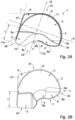
- a prosthesis 1 of the invention for repairing a hernia defect in an inguinal region of the human body.
- the prosthesis 1 shown on Figure 1 is intended to be implanted in the left hand side of a patient body.
- the prosthesis 1 comprises a piece 2 of biocompatible material having a preformed three-dimensional shape.
- On Figure 2A are indicated the medial-lateral axis A and the superior-inferior axis B.
- the preformed piece 2 of Figure 2A has a lateral side 2a, a medial side 2b, a superior side 2c and an inferior side 2d.
- On Figure 2A is shown the front side of the prosthesis 1.
- the piece 2 comprises a first portion 3, which is intended to face the anterior abdominal wall, a second portion 4, which is intended to face the psoas muscle and a third portion 5, which is intended to face the medial inferior area of the inguinal region.
- the piece 2 further comprises a fourth portion 6, intended to ease the alignment of the prosthesis 1 on the Linea Alba.
- the piece 2 does not comprise such fourth portion.
- the first portion 3, second portion 4, third portion 5 and fourth portion 6 are all united to form the preformed three-dimensional shaped piece 2 as a unitary structure.
- the piece 2 has an edge 7 defined by the contour of its preformed three-dimensional shape.
- the first portion 3 forms a partial spherical cap surface 8.
- the partial spherical cap surface 8 is intended to face the anterior abdominal wall once the prosthesis 1 is implanted in the body of a patient.
- the partial spherical cap surface 8 therefore extends in the front direction, namely towards the abdominal wall.
- the partial spherical cap surface 8 is shaped and dimensioned so as to conform to the curved shape of the anterior abdominal wall.
- the partial spherical cap surface 8 is further intended to be positioned adjacent the psoas muscle in an implanted configuration.
- the partial spherical cap surface 8 is derived from a spherical cap from which an inferior part has been removed, the inferior part removed corresponding to the presence of the psoas muscle.
- the partial spherical cap surface 8 may therefore be a spherical cap surface that has been cut in its inferior part along a line delimited by the shape of the psoas muscle. Such a line forms an inferior edge 8a of the partial spherical cap surface 8.
- the spherical cap from which the first portion 3 is formed may be obtained from the cutting of a cap from a sphere having a diameter ranging from about 200 mm to about 220 mm, preferably ranging from about 206 mm to about 215 mm, where the cut cap has a height H, as shown on Figure 4 , ranging from about 15 mm to about 35 mm, preferably from about 20 mm to about 28 mm.
- the partial spherical cap surface 8 forming the first portion 3 may result from the removal of an inferior part of such a spherical cap along a wavy line forming the inferior edge 8a of the partial spherical cap 8 and of the first portion 3.
- the second portion 4 forms a wavy-shaped wall 9 intended to face the psoas muscle once the prosthesis 1 is implanted in the body of a patient.
- the wavy-shaped wall 9 extends from the inferior edge 8a of the first portion 3 and is shaped and dimensioned so as to conform to the shape of the psoas muscle.
- the wavy-shaped wall 9 extends from a lateral side 2a to substantially a medial side 2b of the piece 2.
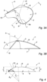
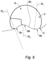
- the wavy-shaped wall 9 includes a surface generated by a generatrix, under the form of a straight line D shown for example on Figures 3A and 3B , following a directrix, under the form of a directing curved line C shown for example on Figure 5 .
- the wavy-shaped wall 9 may comprise a succession of several curves, for example two or three curves, forming a wave.
- the wavy-shaped wall 9 comprises three curves, namely a lateral curve 9a extending substantially in the inferior direction, a central curve 9b extending substantially in the superior direction and a medial curve 9c extending in the inferior direction.
- FIG. 5 is shown a schematic top view of the first portion 3 of Figure 2A showing the directing curved line C including the lateral curve 9a, the central curve 9b and the medial curve 9c and their corresponding radius of curvatures (R1, R2, R3).
- the radius of curvature of the lateral curve 9a may range from about 50 mm to about 55 mm, and may preferably be about 53 mm
- the radius of curvature of the central curve 9b, shown as R2 on Figure 5 may range from about 20 mm to about 35 mm, and may preferably be from about 24 mm to about 31 mm
- the radius of curvature of the medial curve 9c, shown as R3 on Figure 5 may range from about 70 mm to about 90 mm, and may preferably be about 80 mm.
- the wavy-shaped wall 9 is inclined with respect to the direction of the height "h" of the spherical cap 8 of the first portion 3.
- the angle ⁇ formed between the generatrix D of the wavy-shaped wall and the direction "h" of the height of the spherical cap surface 8 of the first portion 3 may range from about 35° to about 50°, preferably from about 40° to about 45°.
- FIG. 2B is shown a second embodiment of the prosthesis 1 of Figure 1 , in which the wavy-shaped wall 9 does not comprise any medial curve.
- the prosthesis of Figure 2B may be designed for cases where the size of the hernia defect in the medial inferior area of the inguinal region requires high coverage from the prosthesis 1.
- the third portion 5 is made larger and replaces the medial curve of the wavy-shaped wall 9.
- the third portion 5 forms an arched part 10.
- the arched part 10 extends longitudinally substantially in the inferior direction from a medial inferior corner 3a of the first portion 3. As better seen on Figure 4 , the arched part 10 extends radially substantially in the front direction.
- the arched part 10 forming the third portion 5 is intended to face the medial inferior area of the inguinal anatomy.
- the radius R4 of the arched part 10 is the radius of the tube from which the arched part derives and is shown in Figure 4 .
- the arched part 10 may have a radius R4 ranging from about 70 mm to about 110 mm, preferably from about 80 mm to about 100 mm.
- the arched part 10 extends circumferentially along a portion of a circle forming an angle ⁇ ranging from about 30° to about 45°, preferably ranging from 33° to 40°.
- the arched part 10 may have a height J ranging from about 20 mm to about 40 mm, preferably from about 21 mm to about 35 mm.
- the arched part 10 may have a length L ranging from about 40 mm to about 60 mm, preferably from about 45 mm to about 53 mm.
- the arched part 10 allows covering the various organs present in the medial inferior area of the inguinal region.
- the shape and dimension of the arched part 10 allow spreading easily the prosthesis 1 without having to tear it or to create specific folds in order to adapt to the unique anatomy of the patient to be treated.
- the height J of the arched part 10 is greater than the height H of the spherical cap of the first portion 3.
- the fourth portion 6 extends from the superior-medial part of the first portion 3.
- the fourth portion 3 forms a triangular part 12 defining a superior-medial corner of the prosthesis 1.
- this corner may form an angle ⁇ , as shown on Figure 2A , ranging from about 100° to about 120°, preferably from about 105° to about 115°, for example of about 110°.
- the triangular part 12 allows the prosthesis 1 to provide an additional reinforcement in the medial superior area of the inguinal region. Such embodiments are particularly suitable when the hernia to be repaired is a direct inguinal hernia.
- the presence of the fourth portion 6 may also help the surgeon positioning optimally the prosthesis 1 by making the medial edge of the prosthesis 1 more visible to the surgeon. The surgeon may then more easily align the medial edge of the prosthesis 1 with the Linea Alba.
- edge 7 extends in the three dimensions of the space. This allows the piece 2 to follow the anatomy of the region to be protected in an optimum way.
- the edge 7 is provided with a reinforcement member 11.
- the reinforcement member 11 forms a non-elastic belt maintaining the prosthesis 1 and helps the handling of the prosthesis 1 while providing a pop-up effect when the prosthesis 1 has been folded on itself, like in a trocar for example.
- the four portions (3, 4, 5, 6) of the prosthesis 1 of Figure 2A are preferably made from a porous textile, in particular a textile showing an elasticity allowing it to be deformed when submitted to an outer pressure and to come back to its initial predetermined three-dimensional shape when said outer pressure is released.
- the reinforcement member 11 may be a fused part of the contour of the textile forming the piece 2, for example obtained by thermal welding. Such a fused part shows a smooth outer shape and is free of any traumatic element. Such a fused part also allows avoiding self-gripping of the textile when the prosthesis is folded on itself during its introduction to the implantation site via a trocar.
- the piece 2 of the prosthesis 1 may be obtained using a compressive thermoforming process : for example a flat textile such as the textile described above is secured between the two parts of a mold having the desired shape for the piece of the prosthesis to be obtained. The whole is heated at temperature of about 140 °C and then cooled down in order to obtain the preformed three-dimensional shaped piece 2.
- the preformed three-dimensional shaped piece thus obtained from said textile shows an elasticity allowing it to be deformed when submitted to an outer pressure and to come back to its initial predetermined three-dimensional shape when said outer pressure is released.
- Fusing the edge of the piece except for the medial inferior part of the edge allows forming a prosthesis capable of spreading out automatically from a trocar and having a good handleability, while showing in its medial inferior part an elasticity enabling the surgeon to conform the prosthesis to the medial inferior area of the inguinal region of the patient being treated.
- the prosthesis of the invention is adapted to be used in the repair of inguinal hernia, such as the direct inguinal hernia, the indirect inguinal hernia and/or the femoral inguinal hernia.
- inguinal hernia such as the direct inguinal hernia, the indirect inguinal hernia and/or the femoral inguinal hernia.
- the prosthesis of the invention allows covering the medial inferior area of the inguinal anatomy, in particular the region around the upper part of the pubic bone, without having to tear or stretch the other parts of the prosthesis or to create additional folds likely to undesirably interfere with the surrounding organs.
Landscapes
- Health & Medical Sciences (AREA)
- Cardiology (AREA)
- Oral & Maxillofacial Surgery (AREA)
- Transplantation (AREA)
- Engineering & Computer Science (AREA)
- Biomedical Technology (AREA)
- Heart & Thoracic Surgery (AREA)
- Vascular Medicine (AREA)
- Life Sciences & Earth Sciences (AREA)
- Animal Behavior & Ethology (AREA)
- General Health & Medical Sciences (AREA)
- Public Health (AREA)
- Veterinary Medicine (AREA)
- Prostheses (AREA)
Description
- The present invention relates to a preformed three-dimensional prosthesis to be used for repair of inguinal hernias.
- In this application, the "medial" end or part of an element of a prosthesis is to be understood as meaning the end or part of the element located in the direction of the median plane of the body when the prosthesis is implanted in the body. The "lateral" end or part of an element of a prosthesis is to be understood as meaning the end or part of the element located in the direction of the outwards lateral plane of the body when the prosthesis is implanted in the body. Likewise, in this application, the "medial direction" is to be understood as meaning the direction towards said median plane and the "lateral direction" is opposite the "medial direction", the medial and lateral directions being aligned on the same axis, the medial-lateral axis. In this application, the "superior" end or part of an element of a prosthesis is to be understood as meaning the end or part of the element located substantially in the direction of the head of the body when the prosthesis is implanted in the body. The "inferior" end or part of an element of a prosthesis is to be understood as meaning the end or part of the element located in the direction of the feet of the body when the prosthesis is implanted in the body. Likewise, in this application, the "superior direction" is to be understood as meaning the direction towards said head and the "inferior direction" is opposite the "superior direction", the superior and inferior directions being aligned on the same axis, the superior-inferior axis. In this application, the "front" end or part of an element of a prosthesis is to be understood as meaning the end or part of the element located substantially in the direction of the front of the body when the prosthesis is implanted in the body. The "rear" end or part of an element of a prosthesis is to be understood as meaning the end or part of the element located in the direction of the back of the body when the prosthesis is implanted in the body. Likewise, in this application, the "front direction" is to be understood as meaning the direction towards said front and the "rear direction" is opposite the "front direction", the front and rear directions being aligned on the same axis, the front-rear axis.
- The abdominal wall in humans is composed of fat and muscles interconnected by fascias. It sometimes happens that a break in continuity occurs in the fascias, allowing part of the peritoneum to slip through and form a sac, or a hernia, containing either fat or part of the intestines. Hernias or incisional hernias (a hernia occurring through a parietal surgical scar) show themselves in the form of a bulge at the surface of the skin and are classed, for example, as umbilical or inguinal hernias, depending on where they are located.
- Wall reinforcement prostheses, for example for the abdominal wall, are widely used in surgery. These prostheses are intended to treat hernias by temporarily or permanently filling a tissue defect. These prostheses are generally made from a biocompatible prosthetic textile and can have a number of shapes, for example rectangular, circular or oval, depending on the anatomical structure to which they are to adapt. Some of these prostheses may show three-dimensional shapes.
- Indeed, when an inguinal hernia is to be treated, it is of particular importance to take into account the anatomy of the inguinal region, in particular the presence of the iliac vessels. In addition, when the patient is a man, the spermatic cord needs to be taken into account while positioning the prosthesis. The various anatomical organs to be taken into account confer to the inguinal region to be treated a three-dimensional shape. In addition, the shape of the inguinal region being asymetric, the shape of a prosthesis intended to be used for treating an inguinal hernia will be dependent on the side (right or left) of the body that is to be treated. In this view, the shape of a prosthesis for treating an inguinal hernia may be defined in relation with the position of the prosthesis once implanted in the body of a patient. For example, in an implanted configuration, a three-dimensional prosthesis for treating an inguinal hernia may comprise a medial part, a lateral part, a superior part, an inferior part, a front part and a rear part as defined above.
- With reference to
Figure 1 , on which are indicated the medial-lateral axis A and the superior-inferior axis B, the anterior retro-parietal space of the inguinal region for the right hand side of a body is shown ; in other words the inguinal region is shown from a view point located in the rear side of the body, for example from the interior of the abdominal cavity but with the peritoneum not shown on the Figure for sake of clarity, and looking towards the front side of the body, in other words in the direction of the abdominal skin of the body. OnFigure 1 , one can see : - in direction of the front side of the body, the rectus abdominis muscles 116 and the transverse muscle 118 forming part of the anterior abdominal wall of the abdomen,
- in the direction of the lateral side of the body, the
psoas muscle 112, the iliac vessels 111, and the elements of thespermatic cord 120, - in the direction of the medial inferior side of the body, the upper part of the
pubic bone 117. - On
Figure 1 , are represented in dotted lines the three locations for potential hernias to occur in the inguinal region : the indirect inguinal hernia, shown as dottedline circle 122 onFigure 1 , occurs mainly in the lateral area of the inguinal region : it is a swelling of the groin caused when a portion of the peritoneum (not shown but located between theFigure 1 and the person looking atFigure 1 ), possibly containing abdominal viscera, passes through the orifice of the inguinal canal. It is necessary to push the peritoneum, and possibly the abdominal viscera, back in the direction of the abdominal cavity, and place a barrier, namely a prosthesis, between the peritoneum and the orifice of the inguinal canal. The direct hernia, shown as dottedline circle 119 onFigure 1 , occurs mainly in the medial area of the inguinal region. The femoral hernia, shown asdotted line circle 121 onFigure 1 , occurs mainly in the medial inferior area of the inguinal region. - It will be noted in
Figure 1 that the elements described above are not all in the same spatial plane, but instead are arranged in an oblique arrangement from a superior-lateral corner to an inferior-medial corner. In the case of an inguinal hernia, the prosthesis implanted after reduction of the hernia must ensure satisfactory covering of the hernia to be treated, either direct, indirect or femoral, by adapting to the contours of the region and by respecting the obliqueness of the inguinal region, if possible without leaving any empty spaces. - When operating by laparoscopy for repairing an inguinal hernia, two routes may be used for bringing the prosthesis to the implantation site, namely the inguinal region. For example, according to a first surgical method, namely the transabdominal preperitoneal route (TAPP), the prosthesis is first conveyed to the abdominal cavity via a trocar; the peritoneum is then open and the prosthesis is brought to the inguinal region through the incision performed through the peritoneum. The peritoneum is closed once the implantation is completed. According to a second surgical method, namely the totally extra-peritoneal route (TEP), the prosthesis is brought to the inguinal region through a trocar directly through the muscles of the abdominal wall, and the peritoneum is not open.
- Because of the obliqueness of the inguinal region and the restricted deployment space, it can prove complicated to deploy the prosthesis and then orient it suitably with respect to the orifice of the inguinal canal or to the other organs to be protected, such as the illiac vessels or the spermatic cord.
- The effectiveness of the prosthesis, hence the ability to minimize the risks of recurrence, depends to a large extent on how well the prosthesis is correctly spread out against the biological tissues of the inguinal region. In the present application, "biological tissues of the inguinal region" are understood as the biological tissues of the organs or elements of the inguinal region that are shown in
Figure 1 and that are intended to be protected from the peritoneum with a view to repairing the hernia, and in particular the anterior muscle wall, the upper part of the pubic bone, the iliac and spermatic vessels, and part of the psoas muscle. - Indeed, prostheses based on a textile are generally flexible. In order to introduce them into a trocar, they are often folded up or rolled to reduce their volume. They therefore tend to form creases when introduced at the implantation site. The spreading out of textile based prosthesis from the trocar is of key importance but can prove difficult.
- Documents
US 6, 723, 133 andUS 6, 740, 122 describes a preformed three-dimensional prosthesis for the repair of inguinal hernia intended to conform to the anatomical shape of the defective wall. The prosthesis described in these patents comprises a plurality of distinctly shaped portions joined to each other, all said shaped portions being configured with a substantially spherical shaped. [Insert page 4a] - As seen above, the anatomy of the inguinal region implies various organs such as muscles having various shapes, vessels, such as the illiac vessels, having various paths, and the pubic bone. In addition, the anatomy of the inguinal region may vary significantly from one human being to the other, in particular in its medial inferior area, in the surroundings of the upper part of the pubic bone. It has been observed that, with some preformed three-dimensional protheses of the prior art, this medial inferior area of the inguinal region is not covered efficiently. In particular, depending on the true anatomy of the patient being treated, the surgeon may need to apply a certain tension on the prosthesis or shift the prosthesis at the moment of positioning the prosthesis, so that a part of the prosthesis be present in the medial inferior area of the inguinal region, namely in the surroundings of the pubic bone. Alternatively the surgeon may need to create folds in the prosthesis so as to conform it to the specific shape of the medial inferior area of the inguinal region he is confronted to in relation with the patient he is treating.
- There is therefore still the need for a prosthesis for repair of inguinal hernias that is based on a preformed three-dimensional piece of biocompatible material intended to globally conform to the anatomical shape of the defective wall in the inguinal region, that is able to be spread out from the trocar and cover efficiently not only said defective wall but also the medial inferior area of the inguinal region, whatever the true anatomical shape of said medial inferior area in the patient to be treated, such that the surgeon does not have to apply specific tensions on the prosthesis or create folds when positioning the prosthesis with respect to the biological tissues.
- The present invention aims to meet such a need.
-
Document EP 3 000 432 A1 describes a prosthesis for hernia repair comprising portions for covering the anterior muscle wall and the psoas muscle. DocumentEP 0 836 838 A1 describes a prosthesis for hernia repair comprising a portion for covering the psoas muscle. DocumentWO95/07666 US2013/0178875 describes a prosthesis for hernia repair. - A first aspect of the invention is an implantable prosthesis for repairing a hernia defect in an inguinal region of a human body delimited by the anterior abdominal wall, the psoas muscle and a medial inferior area of the inguinal anatomy, the prosthesis being according to
claim 1. Further aspects are defined in the dependent claims. - The prosthesis of the invention has a shape allowing it to conform ideally to the anatomy of the inguinal region to be reinforced. In particular, the shape and dimension of the first, second and third portions allow adapting the prostheses to different types of inguinal hernia, such as the direct inguinal hernia, the indirect inguinal hernia and the femoral inguinal hernia.
- Moreover, the presence of the third portion of the prosthesis of the invention and the specific shape of this third portion allow covering the medial inferior area of the inguinal anatomy, in particular the region around the upper part of the pubic bone, without having to tear or stretch the other parts of the prosthesis or to create additional folds likely to undesirably interfere with the surrounding organs. Indeed, the presence of this third portion and its shape allow the surgeon to easily lay the entire prosthesis on each anatomical element to be protected without having to apply a particular compensating force on another part of the prosthesis or to shift the prosthesis. The prosthesis is perfectly spread out in the three dimensions of the inguinal region to be repaired, with no need to create folds in the prosthesis in order to conform it to the shape of the anatomy to be protected. There is no tension created in the prosthesis when it is positioned.
- The prosthesis of the invention is intended to be used for repairing a hernia defect in the inguinal region of a human body. The prosthesis of the invention is particularly adapted for use in laparoscopic surgery, either via the transabdominal preperitoneal route (TAPP) or the totally extra-peritoneal route (TEP).
- The inguinal region may be delimited by the anterior abdominal wall, the psoas muscle and a medial inferior area of the inguinal anatomy, in the surroundings of the pubic bone.
- The prosthesis of the invention comprises a piece of biocompatible material having a preformed three-dimensional shape. The piece of biocompatible material comprises several portions, each having a determined shape, the assembly of these determined shapes forming the preformed three-dimensional shape. The piece of biocompatible material is preferably made as a unitary structure.
- In the present application, "biocompatible" is understood as meaning that the materials having this property can be implanted in the human or animal body.
- Biocompatible materials for forming hernia repair prosthesis are well known in the art. For example, the biocompatible material may comprise a bioresorbable, a non-bioresorbable material and mixtures thereof.
- In the present application, "bioresorbable" or "biodegradable" is understood to mean that the materials having this property are absorbed and/or degraded by the tissues or washed from the implantation site and disappear in vivo after a certain time, which may vary, for example, from a few hours to a few years, depending on the chemical nature of the materials.
- Examples of bioresorbable material suitable for the piece of the prosthesis of the invention can be chosen from among the following bioresorbable materials: polylactic acid (PLA), polycaprolactones (PCL), polydioxanones (PDO), trimethylene carbonates (TMC), polyvinyl alcohol (PVA), polyhydroxyalkanoates (PHA), oxidized cellulose, polyglycolic acid (PGA), polyethylene glycol (PE), copolymers of these materials, and mixtures thereof.
- Examples of non-bioresorbable material suitable for the piece of the prosthesis of the invention can be chosen from among the following non-bioresorbable materials: polypropylenes, polyesters such as polyethylene terephthalates, polyamides, silicones, polyether ether ketone (PEEK), polyarylether ether ketone (PAEK) and mixtures thereof.
- The piece may be made from a preformed sheet of foam, preferably a porous material. Within the meaning of the present application, "porous material" is understood as a material having pores, voids or holes, that are open and are distributed uniformly or irregularly and promote cell colonization and tissue ingrowth. The pores can be present in all types of configurations, for example as spheres, channels, hexagonal forms.
- In embodiments, the piece of biocompatible material comprises a textile, in particular a porous textile. In embodiments, the piece of biocompatible material consists in a textile, for example a porous textile.
- According to the present invention, "textile" is understood as any arrangement or assembly of biocompatible yarns, fibres, filaments and/or multifilaments, for example obtained by knitting, weaving, braiding, or non-woven. Biocompatible textiles, in particular porous textiles, suitable for the repair of a hernia defect are well known in the art.
- The piece of biocompatible material of the prosthesis of the invention comprises a first portion forming a partial spherical cap surface. The partial spherical cap surface is intended to face the anterior abdominal wall. The partial spherical cap surface may extend in the front direction, namely towards the abdominal wall. The partial spherical cap surface is shaped and dimensioned so as to conform to the shape of the anterior abdominal wall. A spherical cap suitable for forming said first portion may derive from a sphere having a diameter ranging from about 200 mm to about 220 mm, preferably ranging from about 206 mm to about 215 mm, said spherical cap having a height ranging from about 15 mm to about 35 mm, preferably from about 20 mm to about 28 mm. The partial spherical cap suitable for obtaining the surface forming the first portion may result from the removal of an inferior part of the spherical cap along a wavy line forming an inferior edge of the first portion.
- The piece of biocompatible material of the prosthesis of the invention comprises a second portion forming a wavy-shaped wall intended to face the psoas muscle. The wavy-shaped wall extends from the inferior edge of the first portion and is shaped and dimensioned so as to conform to the shape of the psoas muscle. In embodiments, the wavy-shaped wall extends substantially from a lateral side to a medial side of the piece of biocompatible material and it includes a surface generated by a generatrix, under the form of a straight line D, following a directrix, under the form of a directing curved line C. In embodiments, the directing curved line C includes at least a lateral curve extending substantially in the inferior direction and at least a central curve, offset in the medial direction with respect to said lateral curve, said central curve extending substantially in the superior direction. In embodiments, the directing curved line C further includes a medial curve extending substantially in the inferior direction. The presence or not of the medial curve may depend on the anatomy of the patient to be treated, in particular on the size of the hernia defect to be repaired.
- For example, the radius of curvature of the lateral curve may range from about 50 mm to about 55 mm, and may preferably be about 53 mm, the radius of curvature of the central curve may range from about 20 mm to about 35 mm, and may preferably be from about 24 mm to about 31 mm, and the radius of curvature of the medial curve, when present, may range from about 70 mm to about 90 mm, and may preferably be about 80 mm.
- The wavy-shaped wall is preferably inclined with respect to the direction of the height of the spherical cap of the first portion. In embodiments, the angle formed between the generatrix of the wavy-shaped wall and the direction of the height of the spherical cap of the first portion may range from about 35° to about 50°, preferably from about 40° to about 45°.
- The piece of biocompatible material further comprises a third portion forming an arched part. The arched part extends longitudinally substantially in the inferior direction from a medial inferior corner of said first portion, said arched part extending radially substantially in the front direction, said third portion being intended to face the medial inferior area of the inguinal anatomy. By "arched part" is meant in the present application an angular section of a tube. The radius of the arched part according to the present application is the radius of the tube from which the arched part derives.
- In embodiments, the arched part has a radius ranging from about 70 mm to about 110 mm, preferably from about 80 mm to about 100 mm. In embodiments, the arched part extends circumferentially along a portion of a circle forming an angle ranging from about 30° to about 45°, preferably ranging from 33° to 40°. The arched part may have a height ranging from about 20 mm to about 40 mm, preferably from about 21 mm to about 35 mm. The arched part may have a length ranging from about 40 mm to about 60 mm, preferably from about 45 mm to about 53 mm.
- The arched part allows covering the various organs present in the medial inferior area of the inguinal region. In particular, the shape and dimension of the arched part allow spreading easily the prosthesis without having to tear it or to create specific folds in order to adapt to the unique anatomy of the patient to be treated.
- In embodiments, the height of the arched part is greater or equal, preferably greater, than the height of the spherical cap of the first portion. The arched part is therefore allowed to cover the medial inferior area of the inguinal region while the spherical cap conforms to the shape of the anterior abdominal wall, without having to apply any specific tension on the prosthesis in the front or rear directions.
- In embodiments, said preformed three-dimensional shape defining an edge of said piece, said edge extends in the three dimensions of the space. The edge of the piece of biocompatible material follows the anatomy of the region to be protected in an optimum way. In particular, thanks to the fact that the edge of the piece extends in the three dimensions of the space, there is no need for the surgeon to create additional folds of the prosthesis in order to conform it to the anatomic shape to be protected.
- In embodiments, said preformed three-dimensional shape defining an edge of said piece, said edge is provided with a reinforcement member at least on a part of its perimeter. The reinforcement member may be made from any biocompatible material and may run continuously or discontinuously along the edge. The reinforcement member may be selected from a moulded material, a wire, a fused textile part and combinations thereof. The reinforcement member is usually provided with a rigidity superior to that of the piece of biocompatible material and may help the handling of the prosthesis during the surgical operation.
- In embodiments, the reinforcement member shows no elasticity along the perimeter of the edge. In other words, if one holds the piece at two points of its edge located substantially on a linear section of said edge, and tries to draw away one point from the other, the reinforcement member will show no elongation between the two points. The reinforcement member may therefore constitute a sort of three-dimensional non-elastic belt maintaining the edge of the piece of biocompatible material, where such belt provides a pop-up effect at the time the prosthesis comes out of a trocar for example. The reinforcement member therefore contributes to a more efficient deployment of the prosthesis when it comes out of the trocar.
- In embodiments, the piece of biocompatible material may show an elasticity allowing it to be deformed when submitted to an outer pressure and to come back to its initial predetermined three-dimensional shape when said outer pressure is released. The surgeon may therefore be able to reduce the global volume of the prosthesis by applying an outer pressure on the prosthesis so as to fold it on itself at the time he introduces the prosthesis in the trocar. When the prosthesis is released from the trocar on the implantation site, namely in the inguinal region, the prosthesis comes naturally to its initial spread out three-dimensional shape.
- A piece of biocompatible material showing such an elasticity may be made from a porous foam.
- Alternatively, some textiles may form biocompatible material showing such an elasticity. A textile suitable for forming the piece of the prosthesis of the invention and showing an elasticity allowing it to be deformed when submitted to an outer pressure and to come back to its initial predetermined three-dimensional shape when said outer pressure is released is described in
US 6, 478, 727 - In embodiments, the reinforcement member comprises a fused part of a contour of the textile forming the piece of biocompatible material. Alternatively, the reinforcement member may consist in a fused part of a contour of said textile. The fused part of the contour of a textile allows forming a reinforcement member having a smooth outer shape and free of any potential traumatic part. The fused part of the contour of a textile further allows forming a reinforcement member showing no elasticity along the perimeter of the edge. The fused part of the contour of the textile also allows avoiding the self-gripping of the textile.
- The contour of the textile may be fused via thermal welding, such as thermal impulse sealing, ultrasonic welding, or induction welding. For example, when thermal impulse sealing is used, the contour of the textile is compressed between two jaws and heated to the melting point of the material forming the textile.
- In embodiments, a medial inferior part of the edge is free of any reinforcement member. The absence of any reinforcement member at the medial inferior part of the edge of the piece of the prosthesis of the invention allows the surgeon to freely direct and position the medial inferior part of the prosthesis in function of the shape of the anatomical organs located in the area of the medial inferior area of the inguinal region. Indeed, it is known that the anatomy of the medial inferior area of the inguinal region varies significantly from one human being to the other. Leaving the medial inferior part of the prosthesis free of any reinforced edge enables the surgeon to adapt the positioning and to customize the fixation of the prosthesis in this area to the true shape of the anatomy encountered for a specific patient. The medial inferior part of the prosthesis is therefore more conformable to the anatomical relief encountered in the treated patient.
- In embodiments, the piece of biocompatible material is a textile showing an elasticity allowing it to be deformed when submitted to an outer pressure and to come back to its initial predetermined three-dimensional shape when said outer pressure is released, having a reinforcement member which is a fused part of said textile, where the medial inferior part of the edge is free of any reinforcement member. Such embodiments allow having a non-elastic belt, formed by the reinforcement member, maintaining the prosthesis on the main part of its edge, while leaving the medial inferior part of the edge preserve its elasticity, coming from the elasticity of the textile it is made of. Such embodiments allow the surgeons to benefit from the elasticity of the medial inferior part of the edge of the textile in order to easily move the prosthesis in the medial inferior area of the inguinal region and to more freely adapt the position of the prosthesis to this specific part of the anatomy, while at the same time having a prosthesis that has enough global rigidity to be manipulated efficiently and to be spread out easily from the trocar.
- In embodiments, the piece further includes a fourth portion extending from the superior-medial part of the first portion, said fourth portion forming a triangular part defining a superior-medial corner of the prosthesis, said corner forming an angle ranging from about 100° to about 120°, preferably from about 105° to about 115°, for example of about 110°. Such a fourth portion allows providing an indication to the surgeon for better positioning the prosthesis. In particular, such a fourth portion helps the surgeon to orientate the prosthesis with regards to the Linea Alba. In addition, the presence of the triangular part forming the fourth portion of the prosthesis allows increasing the surface of the prosthesis in the medial superior area of the inguinal region. Such embodiments are particularly preferred for direct inguinal hernia as they consequently provide additional reinforcement in said medial superior area.
- The preformed three-dimensional shape of the prosthesis of the invention may be obtained using any preforming process known in the art, such as a compressive thermoforming process using a traditional two-parts mold. For example a first part of the mold exhibits an outer surface having the desired shape for the piece of the prosthesis to be obtained and a second part of the mold has a shape similar to that of the first part but recessed into its inner surface. The sheet of biocompatible material, for example a textile, is then secured between the first and the second parts of the mold, the whole being heated at a temperature ranging from about 140°C to about 150°C and further cooled down. The preformed three-dimensional shaped piece thus obtained is removed from the mold.
- The present invention will become clearer from the following description and from the attached drawings, in which:
-
Figure 1 is a schematic view of the inguinal region, -
Figure 2A is a top view of a first embodiment of a prosthesis of the invention, -
Figure 2B is a top view of a second embodiment of the prosthesis of the invention, -
Figures 3A is a side perspective view of the prosthesis ofFigure 2A , -
Figure 3B is a cross section view of the prosthesis ofFigure 3A taken along line I-I, showing the angle between the direction of the height of the spherical cap and the generatrix of the wavy-shaped wall, -
Figure 4 is a side view of the prosthesis ofFigure 2A showing the radius of the arched part, the angle of the portion of a circle on which extends the arched part, and the height of the arched part, -
Figure 5 is a schematic top view of the first portion of the prosthesis ofFigure 2A showing the radius of curvature of the curves of the wavy-shaped wall. - With reference to
Figure 2A , is shown aprosthesis 1 of the invention for repairing a hernia defect in an inguinal region of the human body. Theprosthesis 1 shown onFigure 1 is intended to be implanted in the left hand side of a patient body. Theprosthesis 1 comprises apiece 2 of biocompatible material having a preformed three-dimensional shape. OnFigure 2A are indicated the medial-lateral axis A and the superior-inferior axis B. The preformedpiece 2 ofFigure 2A has alateral side 2a, amedial side 2b, asuperior side 2c and aninferior side 2d. OnFigure 2A is shown the front side of theprosthesis 1. - The
piece 2 comprises afirst portion 3, which is intended to face the anterior abdominal wall, asecond portion 4, which is intended to face the psoas muscle and athird portion 5, which is intended to face the medial inferior area of the inguinal region. In the example shown thepiece 2 further comprises afourth portion 6, intended to ease the alignment of theprosthesis 1 on the Linea Alba. In embodiments not shown, thepiece 2 does not comprise such fourth portion. Thefirst portion 3,second portion 4,third portion 5 andfourth portion 6 are all united to form the preformed three-dimensionalshaped piece 2 as a unitary structure. Thepiece 2 has anedge 7 defined by the contour of its preformed three-dimensional shape. - Each portion (3, 4, 5, 6) of the
piece 2 of biocompatible material will now be described in detail. - The
first portion 3 forms a partialspherical cap surface 8. The partialspherical cap surface 8 is intended to face the anterior abdominal wall once theprosthesis 1 is implanted in the body of a patient. The partialspherical cap surface 8 therefore extends in the front direction, namely towards the abdominal wall. The partialspherical cap surface 8 is shaped and dimensioned so as to conform to the curved shape of the anterior abdominal wall. The partialspherical cap surface 8 is further intended to be positioned adjacent the psoas muscle in an implanted configuration. In this view, the partialspherical cap surface 8 is derived from a spherical cap from which an inferior part has been removed, the inferior part removed corresponding to the presence of the psoas muscle. The partialspherical cap surface 8 may therefore be a spherical cap surface that has been cut in its inferior part along a line delimited by the shape of the psoas muscle. Such a line forms aninferior edge 8a of the partialspherical cap surface 8. - The spherical cap from which the
first portion 3 is formed may be obtained from the cutting of a cap from a sphere having a diameter ranging from about 200 mm to about 220 mm, preferably ranging from about 206 mm to about 215 mm, where the cut cap has a height H, as shown onFigure 4 , ranging from about 15 mm to about 35 mm, preferably from about 20 mm to about 28 mm. The partialspherical cap surface 8 forming thefirst portion 3 may result from the removal of an inferior part of such a spherical cap along a wavy line forming theinferior edge 8a of the partialspherical cap 8 and of thefirst portion 3. - The
second portion 4 forms a wavy-shapedwall 9 intended to face the psoas muscle once theprosthesis 1 is implanted in the body of a patient. The wavy-shapedwall 9 extends from theinferior edge 8a of thefirst portion 3 and is shaped and dimensioned so as to conform to the shape of the psoas muscle. - With reference to
Figure 2A , the wavy-shapedwall 9 extends from alateral side 2a to substantially amedial side 2b of thepiece 2. The wavy-shapedwall 9 includes a surface generated by a generatrix, under the form of a straight line D shown for example onFigures 3A and 3B , following a directrix, under the form of a directing curved line C shown for example onFigure 5 . The wavy-shapedwall 9 may comprise a succession of several curves, for example two or three curves, forming a wave. In the example shown inFigure 2A , the wavy-shapedwall 9 comprises three curves, namely alateral curve 9a extending substantially in the inferior direction, acentral curve 9b extending substantially in the superior direction and amedial curve 9c extending in the inferior direction. - With reference to
Figure 5 , is shown a schematic top view of thefirst portion 3 ofFigure 2A showing the directing curved line C including thelateral curve 9a, thecentral curve 9b and themedial curve 9c and their corresponding radius of curvatures (R1, R2, R3). - For example, the radius of curvature of the
lateral curve 9a, shown as R1 onFigure 5 , may range from about 50 mm to about 55 mm, and may preferably be about 53 mm, the radius of curvature of thecentral curve 9b, shown as R2 onFigure 5 , may range from about 20 mm to about 35 mm, and may preferably be from about 24 mm to about 31 mm, and the radius of curvature of themedial curve 9c, shown as R3 onFigure 5 , may range from about 70 mm to about 90 mm, and may preferably be about 80 mm. - The wavy-shaped
wall 9 is inclined with respect to the direction of the height "h" of thespherical cap 8 of thefirst portion 3. With reference toFigure 3A and 3B , the angle α formed between the generatrix D of the wavy-shaped wall and the direction "h" of the height of thespherical cap surface 8 of thefirst portion 3 may range from about 35° to about 50°, preferably from about 40° to about 45°. - With reference to
Figure 2B , is shown a second embodiment of theprosthesis 1 ofFigure 1 , in which the wavy-shapedwall 9 does not comprise any medial curve. As will appear from the description below, the prosthesis ofFigure 2B may be designed for cases where the size of the hernia defect in the medial inferior area of the inguinal region requires high coverage from theprosthesis 1. Thethird portion 5 is made larger and replaces the medial curve of the wavy-shapedwall 9. - With reference to
Figure 2A , thethird portion 5 forms anarched part 10. Thearched part 10 extends longitudinally substantially in the inferior direction from a medialinferior corner 3a of thefirst portion 3. As better seen onFigure 4 , thearched part 10 extends radially substantially in the front direction. - The
arched part 10 forming thethird portion 5 is intended to face the medial inferior area of the inguinal anatomy. The radius R4 of thearched part 10 is the radius of the tube from which the arched part derives and is shown inFigure 4 . - The
arched part 10 may have a radius R4 ranging from about 70 mm to about 110 mm, preferably from about 80 mm to about 100 mm. - With reference to
Figure 4 , thearched part 10 extends circumferentially along a portion of a circle forming an angle β ranging from about 30° to about 45°, preferably ranging from 33° to 40°. - Still with reference to
Figure 4 , thearched part 10 may have a height J ranging from about 20 mm to about 40 mm, preferably from about 21 mm to about 35 mm. With reference toFigures 2A and 2B , thearched part 10 may have a length L ranging from about 40 mm to about 60 mm, preferably from about 45 mm to about 53 mm. - The
arched part 10 allows covering the various organs present in the medial inferior area of the inguinal region. In particular, the shape and dimension of thearched part 10 allow spreading easily theprosthesis 1 without having to tear it or to create specific folds in order to adapt to the unique anatomy of the patient to be treated. - Still with reference to
Figure 4 , the height J of thearched part 10 is greater than the height H of the spherical cap of thefirst portion 3. Once theprosthesis 1 is implanted, thearched part 10 is thus allowed to cover the medial inferior area of the inguinal region while thespherical cap surface 8 conforms to the shape of the anterior abdominal wall, without having to apply any specific tension on the prosthesis in the front or rear directions. - With reference to
Figure 2A , thefourth portion 6 extends from the superior-medial part of thefirst portion 3. Thefourth portion 3 forms atriangular part 12 defining a superior-medial corner of theprosthesis 1. For example, this corner may form an angle γ, as shown onFigure 2A , ranging from about 100° to about 120°, preferably from about 105° to about 115°, for example of about 110°. Thetriangular part 12 allows theprosthesis 1 to provide an additional reinforcement in the medial superior area of the inguinal region. Such embodiments are particularly suitable when the hernia to be repaired is a direct inguinal hernia. The presence of thefourth portion 6 may also help the surgeon positioning optimally theprosthesis 1 by making the medial edge of theprosthesis 1 more visible to the surgeon. The surgeon may then more easily align the medial edge of theprosthesis 1 with the Linea Alba. - With reference to
Figure 4 , one can see that theedge 7 extends in the three dimensions of the space. This allows thepiece 2 to follow the anatomy of the region to be protected in an optimum way. - With reference to
Figure 2A , theedge 7 is provided with areinforcement member 11. - The
reinforcement member 11 forms a non-elastic belt maintaining theprosthesis 1 and helps the handling of theprosthesis 1 while providing a pop-up effect when theprosthesis 1 has been folded on itself, like in a trocar for example. - On
figure 2A , thereinforcement member 11 runs along a part of theedge 7 while leaving free the medial inferior part of saidedge 7. In particular, thereinforcement member 11 runs from afirst end 11a, approximately located at the level of thecentral curve 9b, to asecond end 11b, approximately located at the medial-inferior corner of thearched part 5, leaving free the medial inferior part of theedge 7. Such an embodiment, where theedge 7 is reinforced except in its medial inferior part, allows obtaining both the required pop-up effect for the deployment of theprosthesis 1 out of a trocar and the possibility to adapt and conform thethird portion 5, namely thearched part 10, to the true anatomy of the medial inferior area of the inguinal region of the patient treated. - The four portions (3, 4, 5, 6) of the
prosthesis 1 ofFigure 2A are made from a biocompatible material. The biocompatible material may comprise a bioresorbable, a non-bioresorbable material and mixtures thereof. - The four portions (3, 4, 5, 6) of the
prosthesis 1 ofFigure 2A are preferably made from a porous textile, in particular a textile showing an elasticity allowing it to be deformed when submitted to an outer pressure and to come back to its initial predetermined three-dimensional shape when said outer pressure is released. Such a textile may for example be a porous knit made of polypropylene monofilament yarn having a diameter ranging from about 0.12 mm to about 0.25 mm, preferably from about 0.15 mm to about and 0.20 mm, for example of about 0.18 mm, and threaded one full one empty in two guide bars according to the following knitting pattern according to ISO 11676 standard, publication 2014 : Bar I : 3-2/2-1/0-1//, Bar II : 0-1/1-2/3-2//. For example, the number of stitches per centimeter for such a porous knit may vary from about 7 to 15, preferably from about 10 to 12. - The
reinforcement member 11 may be a fused part of the contour of the textile forming thepiece 2, for example obtained by thermal welding. Such a fused part shows a smooth outer shape and is free of any traumatic element. Such a fused part also allows avoiding self-gripping of the textile when the prosthesis is folded on itself during its introduction to the implantation site via a trocar. - When the textile is a porous knit obtained from polypropylene monofilament as described above, the reinforcement member may be obtained by fusing the contour of the textile as follows : the contour of the textile is compressed between two jaws and heated to about 240°C which is the melting point of polypropylene.
- The
piece 2 of theprosthesis 1 may be obtained using a compressive thermoforming process : for example a flat textile such as the textile described above is secured between the two parts of a mold having the desired shape for the piece of the prosthesis to be obtained. The whole is heated at temperature of about 140 °C and then cooled down in order to obtain the preformed three-dimensionalshaped piece 2. The preformed three-dimensional shaped piece thus obtained from said textile shows an elasticity allowing it to be deformed when submitted to an outer pressure and to come back to its initial predetermined three-dimensional shape when said outer pressure is released. Fusing the edge of the piece except for the medial inferior part of the edge allows forming a prosthesis capable of spreading out automatically from a trocar and having a good handleability, while showing in its medial inferior part an elasticity enabling the surgeon to conform the prosthesis to the medial inferior area of the inguinal region of the patient being treated. - The prosthesis of the invention is adapted to be used in the repair of inguinal hernia, such as the direct inguinal hernia, the indirect inguinal hernia and/or the femoral inguinal hernia. In particular, the prosthesis of the invention allows covering the medial inferior area of the inguinal anatomy, in particular the region around the upper part of the pubic bone, without having to tear or stretch the other parts of the prosthesis or to create additional folds likely to undesirably interfere with the surrounding organs.
Claims (21)
- An implantable prosthesis (1) for repairing a hernia defect in an inguinal region of a human body delimited by the anterior abdominal wall, the psoas muscle and a medial inferior area of the inguinal anatomy, the prosthesis comprising :a piece (2) of biocompatible material having a preformed three-dimensional shape, the piece (2) having a lateral side (2a) and a medial side (2b) located in opposite directions on a medial-lateral axis (A) and a superior side (2c) and an inferior side (2d) located in opposite directions on a superior-inferior axis (B), the inferior direction corresponding to a direction toward feet of a body when the implantable prosthesis (1) is implanted in the body, the piece including :- a first portion (3) , configured to face the anterior abdominal wall, said first portion forming a partial spherical cap surface (8) shaped and dimensioned so as to substantially conform to the shape of the anterior abdominal wall, said first portion having an inferior edge (8a) extending along a wavy line from the lateral side (2a) to substantially the medial side (2b) and defining a medial inferior corner (3a) of the first portion,- a second portion (4), configured to face the psoas muscle, said second portion extending from the inferior edge (8a) of said first portion (3) and forming a wavy-shaped wall (9) extending from the lateral side (2a) to substantially the medial side (2b) of the piece (2), shaped and dimensioned so as to substantially conform to the shape of the psoas muscle,- a third portion (5) forming an arched part (10), said arched part extending longitudinally substantially in the inferior direction from a medial part of the inferior edge (8a) of said first portion (3) including said medial inferior corner (3a) of said first portion, said arched part extending radially substantially in the front direction, said arched part being shaped so as to cover the organs present in the inferior area of the inguinal region, said third portion being configured to face the medial inferior area of the inguinal anatomy.
- The prosthesis (1) of claim 1, wherein said preformed three-dimensional shape defining an edge (7) of said piece, said edge extends in the three dimensions of the space.
- The prosthesis (1) of claim 1 or 2, wherein said preformed three-dimensional shape defining an edge (7) of said piece, said edge is provided with a reinforcement member (11) at least on a part of its perimeter.
- The prosthesis (1) of claim 3, wherein a medial inferior part of the edge (7) is free of any reinforcement member (11).
- The prosthesis (1) of any one of claims 1-4, wherein the piece (2) of biocompatible material comprises a textile.
- The prosthesis (1) of claim 5, wherein the reinforcement member (11) comprises a fused part of a contour of said textile.
- The prosthesis (1) of any one of claims 3-6, wherein said reinforcement member shows no elasticity along the perimeter of the edge.
- The prosthesis (1) of any one of claims 1-7, wherein the piece (2) of biocompatible material shows an elasticity allowing it to be deformed when submitted to an outer pressure and to come back to its initial predetermined three-dimensional shape when said outer pressure is released.
- The prosthesis (1) of any one of claims 1-8, wherein the spherical cap of said first portion (3) derives from a sphere having a diameter ranging from about 200 mm to about 220 mm, preferably ranging from about 206 mm to about 215 mm, said spherical cap having a height (H) ranging from about 15 mm to about 35 mm, preferably from about 20 mm to about 28 mm.
- The prosthesis (1) of any one of claims 1 to 9, wherein said wavy-shaped wall includes a surface generated by a generatrix, under the form of a straight line D, following a directrix, under the form of a directing curved line C.
- The prosthesis of claim 10, wherein said directing curved line C includes at least a lateral curve (9a) extending substantially in the inferior direction and at least a central curve (9b), offset in the medial direction with respect to said lateral curve, said central curve extending substantially in the superior direction.
- The prosthesis (1) of claim 11, wherein the radius of curvature (R1) of the lateral curve (9a) ranges from about 50 mm to about 55 mm, is preferably about 53 mm, the radius of curvature (R2) of the central curve (9b) ranges from about 20 mm to about 35 mm, preferably ranges from about 24 mm to about 31 mm.
- The prosthesis of claim 11 or 12, wherein the directing curved line C further includes a medial curve (9c) extending in the inferior direction.
- The prosthesis of claim 13, wherein the radius of curvature (R3) of the medial curve (9c) ranges from about 70 mm to about 90 mm, is preferably about 80 mm.
- The prosthesis (1) of any one of claims 10-14, wherein the angle (α) formed between the generatrix (D) of the wavy-shaped wall and the direction "h" of the height of the spherical cap of the first portion (3) ranges from about 35° to about 50°, preferably from about 40° to about 45°.
- The prosthesis (1) of any one of claims 1-15, wherein the arched part (10) has a radius (R4) ranging from about 70 mm to about 110 mm, preferably from about 80 mm to about 100 mm.
- The prosthesis (1) of any one of claims 1-16, wherein the arched part (10) extends circumferentially along a portion of a circle forming an angle (β) ranging from about 30° to about 45°, preferably ranging from 33° to 40°.
- The prosthesis (1) of any one of claims 1-17, wherein the arched part (10) has a height (J) ranging from about 20 mm to about 40 mm, preferably from about 21 mm to about 35 mm.
- The prosthesis (1) of any one of claims 1-18, wherein the arched part (10) has a length (L) ranging from about 40 mm to about 60 mm, preferably from about 45 mm to about 53 mm.
- The prosthesis (1) of any one of claims 1-19, wherein the piece (2) further includes a fourth portion (6) extending from the superior-medial part of the first portion, said fourth portion forming a triangular part (12) defining a superior-medial corner of the prosthesis, said corner forming an angle (γ) ranging from about 100° to about 120°, preferably from about 105° to about 115°, for example of about 110°.
- The prosthesis (1) of claims 9 and 18, wherein the height (J) of the arched part (10) is greater or equal, preferably greater, than the height (H) of the spherical cap of the first portion (3).
Priority Applications (7)
| Application Number | Priority Date | Filing Date | Title |
|---|---|---|---|
| EP17305489.1A EP3398554B1 (en) | 2017-05-02 | 2017-05-02 | Prosthesis for inguinal hernia repair |
| AU2018202680A AU2018202680A1 (en) | 2017-05-02 | 2018-04-18 | Prosthesis for inguinal hernia repair |
| CN201810386483.1A CN108784886B (en) | 2017-05-02 | 2018-04-26 | Prosthesis for inguinal hernia repair |
| US15/965,826 US10675137B2 (en) | 2017-05-02 | 2018-04-27 | Prosthesis for inguinal hernia repair |
| CA3003189A CA3003189A1 (en) | 2017-05-02 | 2018-04-30 | Prosthesis for inguinal hernia repair |
| US16/895,888 US11672636B2 (en) | 2017-05-02 | 2020-06-08 | Prosthesis for inguinal hernia repair |
| US18/205,485 US12496178B2 (en) | 2017-05-02 | 2023-06-02 | Prosthesis for inguinal hernia repair |
Applications Claiming Priority (1)
| Application Number | Priority Date | Filing Date | Title |
|---|---|---|---|
| EP17305489.1A EP3398554B1 (en) | 2017-05-02 | 2017-05-02 | Prosthesis for inguinal hernia repair |
Publications (2)
| Publication Number | Publication Date |
|---|---|
| EP3398554A1 EP3398554A1 (en) | 2018-11-07 |
| EP3398554B1 true EP3398554B1 (en) | 2025-06-25 |
Family
ID=58701574
Family Applications (1)
| Application Number | Title | Priority Date | Filing Date |
|---|---|---|---|
| EP17305489.1A Active EP3398554B1 (en) | 2017-05-02 | 2017-05-02 | Prosthesis for inguinal hernia repair |
Country Status (5)
| Country | Link |
|---|---|
| US (3) | US10675137B2 (en) |
| EP (1) | EP3398554B1 (en) |
| CN (1) | CN108784886B (en) |
| AU (1) | AU2018202680A1 (en) |
| CA (1) | CA3003189A1 (en) |
Families Citing this family (7)
| Publication number | Priority date | Publication date | Assignee | Title |
|---|---|---|---|---|
| EP3337427A1 (en) * | 2015-08-21 | 2018-06-27 | Lifecell Corporation | Breast treatment device |
| EP3398554B1 (en) * | 2017-05-02 | 2025-06-25 | Sofradim Production | Prosthesis for inguinal hernia repair |
| CA3079532A1 (en) * | 2017-10-19 | 2019-04-25 | C.R.Bard, Inc. | Self-gripping hernia prosthesis |
| US12064330B2 (en) * | 2020-04-28 | 2024-08-20 | Covidien Lp | Implantable prothesis for minimally invasive hernia repair |
| US11413129B2 (en) * | 2020-06-19 | 2022-08-16 | Davol Inc. | Implantable prosthesis |
| CN114795573B (en) * | 2021-01-27 | 2024-03-19 | 心宜医疗器械(深圳)有限公司 | 3D inguinal hernia repair patch |
| EP4410237A1 (en) | 2023-02-03 | 2024-08-07 | Sofradim Production | Composite implant including a three-dimensional mesh and a foam and methods associated therewith |
Family Cites Families (530)
| Publication number | Priority date | Publication date | Assignee | Title |
|---|---|---|---|---|
| US3124136A (en) | 1964-03-10 | Method of repairing body tissue | ||
| US3118294A (en) | 1964-01-21 | Method for manufacturing knitted nets and products | ||
| US1187158A (en) | 1915-02-18 | 1916-06-13 | Simon Friedberger | Twisted net fabric. |
| US3007531A (en) | 1958-07-17 | 1961-11-07 | Deere & Co | Plow reversible by vane type hydraulic motor |
| US3054406A (en) | 1958-10-17 | 1962-09-18 | Phillips Petroleum Co | Surgical mesh |
| US3364200A (en) | 1960-03-28 | 1968-01-16 | Johnson & Johnson | Oxidized cellulose product and method for preparing the same |
| US3320649A (en) | 1962-10-23 | 1967-05-23 | Naimer Jack | Methods of making separable fastening fabrics |
| US3276448A (en) | 1962-12-14 | 1966-10-04 | Ethicon Inc | Collagen coated fabric prosthesis |
| US3272204A (en) | 1965-09-22 | 1966-09-13 | Ethicon Inc | Absorbable collagen prosthetic implant with non-absorbable reinforcing strands |
| SE344275B (en) | 1966-02-10 | 1972-04-10 | R Gruenert | |
| US3570482A (en) | 1968-12-09 | 1971-03-16 | Fujiboseki Kk | Elastic surgical bandage |
| US3718725A (en) | 1970-11-17 | 1973-02-27 | Int Knitlock Corp | Method for making hook fabric material for fasteners |
| IT993386B (en) | 1973-09-24 | 1975-09-30 | Nannini G E C Sas | PROCEDURE FOR MAKING A LACE IN A RASCHEL TYPE LOOM AND LACE OBTAINED |
| DE2461370A1 (en) | 1974-01-02 | 1975-07-03 | Sauvage Lester R | POROESE VASCULAR PROSTHESIS |
| US4006747A (en) | 1975-04-23 | 1977-02-08 | Ethicon, Inc. | Surgical method |
| US4060081A (en) | 1975-07-15 | 1977-11-29 | Massachusetts Institute Of Technology | Multilayer membrane useful as synthetic skin |
| US4193137A (en) | 1977-05-06 | 1980-03-18 | Meadox Medicals, Inc. | Warp-knitted double-velour prosthesis |
| US4294241A (en) | 1977-06-09 | 1981-10-13 | Teruo Miyata | Collagen skin dressing |
| US4173131A (en) | 1977-08-30 | 1979-11-06 | The Kendall Co. | Porous elastic bandage |
| US4307717A (en) | 1977-11-07 | 1981-12-29 | Lectec Corporation | Sterile improved bandage containing a medicament |
| US4248064A (en) | 1979-02-14 | 1981-02-03 | Stedman Corporation | Lock-stitch knitted elastic fabric |
| BR8000993A (en) | 1979-02-19 | 1980-10-29 | Takeda Lace | TOTALLY URIDID STRIP AND TISSUE AND PROCESS FOR ITS MANUFACTURING |
| US4307496A (en) | 1979-02-19 | 1981-12-29 | Takeda Lace Co., Ltd. | Warp-knitted lace strip, material fabric, and manufacturing method thereof |
| JPS6027281B2 (en) | 1979-05-09 | 1985-06-28 | ワイケイケイ株式会社 | Method for manufacturing velvet fastener tape |
| US4476697A (en) | 1980-04-21 | 1984-10-16 | Karl Otto Braun Kg | Wound dressing |
| DE3042860A1 (en) | 1980-11-13 | 1982-06-09 | Heyl & Co Chemisch-Pharmazeutische Fabrik, 1000 Berlin | COLLAGEN PREPARATIONS, METHODS FOR THEIR PRODUCTION AND THEIR USE IN HUMAN AND VETERINE MEDICINE |
| US4591501A (en) | 1981-04-13 | 1986-05-27 | Seton Company | Cosmetic and pharmaceutical sheet material containing polypeptides |
| FR2516927B1 (en) | 1981-11-26 | 1986-05-23 | Merieux Fond | PROCESS FOR THE INDUSTRIAL PREPARATION OF COLLAGENIC MATERIALS FROM HUMAN PLACENTARY TISSUES, HUMAN COLLAGENIC MATERIALS OBTAINED, THEIR APPLICATION AS BIOMATERIALS |
| US4925294A (en) | 1986-12-17 | 1990-05-15 | Geshwind David M | Method to convert two dimensional motion pictures for three-dimensional systems |
| JPS6014861A (en) | 1983-07-05 | 1985-01-25 | 株式会社日本メデイカル・サプライ | Adhesion preventing material |
| US4487865A (en) | 1983-12-15 | 1984-12-11 | Biomatrix, Inc. | Polymeric articles modified with hyaluronate |
| US4500676A (en) | 1983-12-15 | 1985-02-19 | Biomatrix, Inc. | Hyaluronate modified polymeric articles |
| FR2559780B1 (en) | 1984-02-21 | 1990-05-04 | Tech Cuir Centre | IMPLANTABLE BIOCOMPATIBLE COLLAGEN-BASED SYSTEMS FOR CELL STORAGE AND / OR CULTURE AND / OR CONTROLLED RELEASE OF ACTIVE INGREDIENTS |
| IL74715A0 (en) | 1984-03-27 | 1985-06-30 | Univ New Jersey Med | Biodegradable matrix and methods for producing same |
| US4837285A (en) | 1984-03-27 | 1989-06-06 | Medimatrix | Collagen matrix beads for soft tissue repair |
| FR2577807B1 (en) | 1985-02-22 | 1993-12-03 | Ethnor | ABSORBABLE COMPOSITE SURGICAL MATERIAL, PREPARATION METHOD, RESORBABLE PROSTHESIS MADE FROM SUCH MATERIAL AND USE OF SUCH A PROSTHESIS |
| US4631932A (en) | 1985-05-15 | 1986-12-30 | S.R.C. Textiles, Inc. | Knitted waistband curl-preventing strip |
| US5720981A (en) | 1985-08-14 | 1998-02-24 | Sloan-Kettering Institute For Cancer Research | Epidermal cell extracts and method to enhance wound healing and regenerate epidermis |
| US5002551A (en) | 1985-08-22 | 1991-03-26 | Johnson & Johnson Medical, Inc. | Method and material for prevention of surgical adhesions |
| JPH0235207Y2 (en) | 1985-10-23 | 1990-09-25 | ||
| EP0228203B1 (en) | 1985-12-05 | 1990-07-25 | Sakae Lace Co. Ltd. | Warp knitted lace fabrics |
| US4792336A (en) | 1986-03-03 | 1988-12-20 | American Cyanamid Company | Flat braided ligament or tendon implant device having texturized yarns |
| US4769038A (en) | 1986-03-18 | 1988-09-06 | C. R. Bard, Inc. | Prostheses and techniques and repair of inguinal and femoral hernias |
| GB8611129D0 (en) | 1986-05-07 | 1986-06-11 | Annis D | Prosthetic materials |
| DE3619197A1 (en) | 1986-06-07 | 1987-12-10 | Ethicon Gmbh | UPHOLSTERY IMPLANT |
| FR2601371B1 (en) | 1986-07-11 | 1989-05-12 | Merieux Inst | PROCESS FOR TREATING COLLAGEN WITH A VIEW TO, IN PARTICULAR, FACILITATING CROSS-LINKING AND COLLAGEN OBTAINED BY APPLICATION OF SAID PROCESS |
| US4854316A (en) | 1986-10-03 | 1989-08-08 | Davis Emsley A | Apparatus and method for repairing and preventing para-stomal hernias |
| DE3633974A1 (en) | 1986-10-06 | 1988-04-21 | Bleier Waldemar | LEAD CLIPS |
| US4759354A (en) | 1986-11-26 | 1988-07-26 | The Kendall Company | Wound dressing |
| IT1202456B (en) | 1987-01-30 | 1989-02-09 | Ausonia Spa | TEXTILE MANUFACTURE FOR CONTACT CLOSURE AND METHOD AND EQUIPMENT FOR ITS PRODUCTION |
| US4813942A (en) | 1987-03-17 | 1989-03-21 | Bioderm, Inc. | Three step wound treatment method and dressing therefor |
| FR2612392A1 (en) | 1987-03-19 | 1988-09-23 | Audion Michel | Interrupted biodegradable composites of variable strength |
| US6174999B1 (en) | 1987-09-18 | 2001-01-16 | Genzyme Corporation | Water insoluble derivatives of polyanionic polysaccharides |
| US4937270A (en) | 1987-09-18 | 1990-06-26 | Genzyme Corporation | Water insoluble derivatives of hyaluronic acid |
| US5015584A (en) | 1987-10-14 | 1991-05-14 | Board Of Regents, The University Of Texas System | Epidermal graft system |
| BR8705600A (en) | 1987-10-15 | 1988-08-02 | Ricardo Alfredo Bustos | BREAST PROSTHESIS |
| US4976737A (en) | 1988-01-19 | 1990-12-11 | Research And Education Institute, Inc. | Bone reconstruction |
| US5350583A (en) | 1988-03-09 | 1994-09-27 | Terumo Kabushiki Kaisha | Cell-penetrable medical material and artificial skin |
| WO1989008465A1 (en) | 1988-03-09 | 1989-09-21 | Terumo Kabushiki Kaisha | Medical material permitting cells to enter thereinto and artificial skin |
| FR2628634B1 (en) | 1988-03-15 | 1990-07-13 | Imedex | VISCERAL SURGERY PATCH |
| US5201745A (en) | 1988-03-15 | 1993-04-13 | Imedex | Visceral surgery patch |
| US4950483A (en) | 1988-06-30 | 1990-08-21 | Collagen Corporation | Collagen wound healing matrices and process for their production |
| US4948540A (en) | 1988-08-01 | 1990-08-14 | Semex Medical, Inc. | Method of preparing collagen dressing sheet material |
| US5304595A (en) | 1988-11-21 | 1994-04-19 | Collagen Corporation | Collagen-polymer conjugates |
| US5306500A (en) | 1988-11-21 | 1994-04-26 | Collagen Corporation | Method of augmenting tissue with collagen-polymer conjugates |
| US5162430A (en) | 1988-11-21 | 1992-11-10 | Collagen Corporation | Collagen-polymer conjugates |
| US5800541A (en) | 1988-11-21 | 1998-09-01 | Collagen Corporation | Collagen-synthetic polymer matrices prepared using a multiple step reaction |
| US5614587A (en) | 1988-11-21 | 1997-03-25 | Collagen Corporation | Collagen-based bioadhesive compositions |
| ZA899326B (en) | 1988-12-07 | 1991-08-28 | Johnson & Johnson Patient Care | Low molecular weight heparin,heparinoid and hexuronyl hexosaminoglycan sulfate containing adhesion prevention barrier and process |
| US5171273A (en) | 1989-01-13 | 1992-12-15 | University Of Medicine And Dentistry Of New Jersey | Synthetic collagen orthopaedic structures such as grafts, tendons and other structures |
| FR2641692A1 (en) | 1989-01-17 | 1990-07-20 | Nippon Zeon Co | Plug for closing an opening for a medical application, and device for the closure plug making use thereof |
| DK177289D0 (en) | 1989-04-13 | 1989-04-13 | Coloplast As | bREAST PROSTHESIS |
| US5441508A (en) | 1989-04-27 | 1995-08-15 | Gazielly; Dominique | Reinforcement and supporting device for the rotator cuff of a shoulder joint of a person |
| FR2646343B1 (en) | 1989-04-27 | 1991-12-20 | Gazielly Dominique | DEVICE FOR REINFORCING AND SUPPORTING THE HAIR OF THE ROTATORS OF AN INDIVIDUAL SHOULDER JOINT |
| JPH0332677A (en) | 1989-06-30 | 1991-02-13 | Yoshihiko Shimizu | Vein closing material for imparting phlogistic and clotting property |
| JPH0752229Y2 (en) | 1989-08-07 | 1995-11-29 | 株式会社明治ゴム化成 | Drainage mass |
| US5196185A (en) | 1989-09-11 | 1993-03-23 | Micro-Collagen Pharmaceutics, Ltd. | Collagen-based wound dressing and method for applying same |
| IL95429A (en) | 1989-09-15 | 1997-09-30 | Organogenesis | Living tissue equivalents comprising hydrated collagen lattice and a collagen gel and their production |
| US5201764A (en) | 1990-02-28 | 1993-04-13 | Autogenesis Technologies, Inc. | Biologically compatible collagenous reaction product and articles useful as medical implants produced therefrom |
| US5256418A (en) | 1990-04-06 | 1993-10-26 | Organogenesis, Inc. | Collagen constructs |
| FI924773L (en) | 1990-04-24 | 1992-10-21 | Mark Eisenberg | COMPOSITION SUBSTITUTER AV LEVANDE HUD |
| US5116357A (en) | 1990-10-11 | 1992-05-26 | Eberbach Mark A | Hernia plug and introducer apparatus |
| US5141515A (en) | 1990-10-11 | 1992-08-25 | Eberbach Mark A | Apparatus and methods for repairing hernias |
| US6559119B1 (en) | 1990-11-27 | 2003-05-06 | Loyola University Of Chicago | Method of preparing a tissue sealant-treated biomedical material |
| US6197325B1 (en) | 1990-11-27 | 2001-03-06 | The American National Red Cross | Supplemented and unsupplemented tissue sealants, methods of their production and use |
| US5206028A (en) | 1991-02-11 | 1993-04-27 | Li Shu Tung | Dense collagen membrane matrices for medical uses |
| CA2060223C (en) | 1991-02-12 | 1999-07-20 | Clarence C. Lee | Injectable medical lubricating fluid composition and method of use |
| US5690675A (en) | 1991-02-13 | 1997-11-25 | Fusion Medical Technologies, Inc. | Methods for sealing of staples and other fasteners in tissue |
| US5749895A (en) | 1991-02-13 | 1998-05-12 | Fusion Medical Technologies, Inc. | Method for bonding or fusion of biological tissue and material |
| WO1992015262A1 (en) | 1991-03-06 | 1992-09-17 | Aircast, Inc. | Injection-molded orthopedic device and method |
| US5254133A (en) | 1991-04-24 | 1993-10-19 | Seid Arnold S | Surgical implantation device and related method of use |
| CA2109085C (en) | 1991-04-25 | 2003-03-11 | Keith E. Dionne | Implantable biocompatible immunoisolatory vehicle for delivery of selected therapeutic products |
| US5785983A (en) | 1991-05-23 | 1998-07-28 | Euroresearch Srl | Non-porous collagen sheet for therapeutic use, and the method and apparatus for preparing it |
| US5605938A (en) | 1991-05-31 | 1997-02-25 | Gliatech, Inc. | Methods and compositions for inhibition of cell invasion and fibrosis using dextran sulfate |
| FR2679778B1 (en) | 1991-08-02 | 1995-07-07 | Coletica | USE OF CROLAGEN CROSSLINKED BY A CROSSLINKING AGENT FOR THE MANUFACTURE OF A SLOW RESORPTIVE, BIOCOMPATIBLE, SUTURABLE MEMBRANE, AS WELL AS SUCH A MEMBRANE. |
| US5282829A (en) | 1991-08-15 | 1994-02-01 | United States Surgical Corporation | Hollow body implants |
| FR2682284A1 (en) | 1991-10-14 | 1993-04-16 | Dessapt Bernard | Mammary prosthesis |
| CA2082090C (en) | 1991-11-05 | 2004-04-27 | Jack Fagan | Improved occluder for repair of cardiac and vascular defects |
| US5258000A (en) | 1991-11-25 | 1993-11-02 | Cook Incorporated | Tissue aperture repair device |
| DE69201633T2 (en) | 1991-11-25 | 1995-07-06 | Cook Inc | Device for restoring a tissue opening. |
| DK168419B1 (en) | 1991-11-25 | 1994-03-28 | Cook Inc A Cook Group Company | Abdominal wall support device and apparatus for insertion thereof |
| US5439467A (en) | 1991-12-03 | 1995-08-08 | Vesica Medical, Inc. | Suture passer |
| US5147374A (en) | 1991-12-05 | 1992-09-15 | Alfredo Fernandez | Prosthetic mesh patch for hernia repair |
| WO1993010731A1 (en) | 1991-12-06 | 1993-06-10 | Kensey Nash Corporation | Pads, methods of making, and methods of use for wound dressing, surgical reinforcement and hemostasis promotion |
| US5176692A (en) | 1991-12-09 | 1993-01-05 | Wilk Peter J | Method and surgical instrument for repairing hernia |
| IT1254170B (en) | 1991-12-18 | 1995-09-11 | Mini Ricerca Scient Tecnolog | COMPOSITE MEMBRANES FOR GUIDED REGENERATION OF FABRICS |
| US5376376A (en) | 1992-01-13 | 1994-12-27 | Li; Shu-Tung | Resorbable vascular wound dressings |
| USRE36370E (en) | 1992-01-13 | 1999-11-02 | Li; Shu-Tung | Resorbable vascular wound dressings |
| ES2133382T3 (en) | 1992-01-21 | 1999-09-16 | Univ Minnesota | DEVICE FOR THE CLOSURE OF SEPTAL DEFECTS. |
| FR2686612B1 (en) | 1992-01-24 | 1994-04-08 | Fournier Sca Laboratoires | PROCESS FOR THE PREPARATION OF COLLAGEN FIBERS. |
| JPH07503869A (en) | 1992-02-14 | 1995-04-27 | ボード・オヴ・リージェンツ,ザ・ユニヴァーシティ・オヴ・テキサス・システム | Multiphasic bioerodible implant materials or carriers and methods of manufacture and use thereof |
| CA2090000A1 (en) | 1992-02-24 | 1993-08-25 | H. Jonathan Tovey | Articulating mesh deployment apparatus |
| US5480644A (en) | 1992-02-28 | 1996-01-02 | Jsf Consultants Ltd. | Use of injectable biomaterials for the repair and augmentation of the anal sphincters |
| FR2688222B1 (en) | 1992-03-03 | 1995-05-19 | Univ Picardie | POLYMERIC COMPOUNDS OF GLUCURONIC ACID, PROCESS FOR THE PREPARATION THEREOF, AND USE IN PARTICULAR AS GELIFYING, THICKENING, MOISTURIZING, STABILIZING, CHELATING OR FLOCCULATING MEDIA. |
| WO1993017635A1 (en) | 1992-03-04 | 1993-09-16 | C.R. Bard, Inc. | Composite prosthesis and method for limiting the incidence of postoperative adhesions |
| US5217493A (en) | 1992-03-11 | 1993-06-08 | Board Of Regents, The University Of Texas System | Antibacterial coated medical implants |
| GB9206504D0 (en) | 1992-03-25 | 1992-05-06 | Jevco Ltd | Heteromorphic sponges as wound implants |
| GB9206509D0 (en) | 1992-03-25 | 1992-05-06 | Jevco Ltd | Heteromorphic sponges containing active agents |
| IL105529A0 (en) | 1992-05-01 | 1993-08-18 | Amgen Inc | Collagen-containing sponges as drug delivery for proteins |
| US5456711A (en) | 1992-05-15 | 1995-10-10 | Intervascular Inc. | Warp knitted carotid patch having finished selvedged edges |
| US5766246A (en) | 1992-05-20 | 1998-06-16 | C. R. Bard, Inc. | Implantable prosthesis and method and apparatus for loading and delivering an implantable prothesis |
| US6312442B1 (en) | 1992-06-02 | 2001-11-06 | General Surgical Innovations, Inc. | Method for developing an anatomic space for laparoscopic hernia repair |
| US5428022A (en) | 1992-07-29 | 1995-06-27 | Collagen Corporation | Composition of low type III content human placental collagen |
| US5339657A (en) | 1992-09-01 | 1994-08-23 | Mcmurray Fabrics, Inc. | Net having different size openings and method of making |
| WO1994006460A1 (en) | 1992-09-21 | 1994-03-31 | Vitaphore Corporation | Embolization plugs for blood vessels |
| CZ281454B6 (en) | 1992-11-23 | 1996-10-16 | Milan Mudr. Csc. Krajíček | Aid for non-surgical closing of a hole in a vessel wall |
| US5743917A (en) | 1993-01-13 | 1998-04-28 | Saxon; Allen | Prosthesis for the repair of soft tissue defects |
| CA2114282A1 (en) | 1993-01-28 | 1994-07-29 | Lothar Schilder | Multi-layered implant |
| US5667839A (en) | 1993-01-28 | 1997-09-16 | Collagen Corporation | Human recombinant collagen in the milk of transgenic animals |
| US6653450B1 (en) | 1993-01-28 | 2003-11-25 | Cohesion Technologies, Inc. | Mutated recombinant collagens |
| US5356432B1 (en) | 1993-02-05 | 1997-02-04 | Bard Inc C R | Implantable mesh prosthesis and method for repairing muscle or tissue wall defects |
| US5368602A (en) | 1993-02-11 | 1994-11-29 | De La Torre; Roger A. | Surgical mesh with semi-rigid border members |
| DE69322370C5 (en) | 1993-02-18 | 2009-01-08 | Ethicon Endo-Surgery, Inc., Cincinnati | Laparoscopic adjustable gastric band |
| US5433996A (en) | 1993-02-18 | 1995-07-18 | W. L. Gore & Associates, Inc. | Laminated patch tissue repair sheet material |
| US6015844A (en) | 1993-03-22 | 2000-01-18 | Johnson & Johnson Medical, Inc. | Composite surgical material |
| US6001895A (en) | 1993-03-22 | 1999-12-14 | Johnson & Johnson Medical, Inc. | Composite surgical material |
| US5565210A (en) | 1993-03-22 | 1996-10-15 | Johnson & Johnson Medical, Inc. | Bioabsorbable wound implant materials |
| US5942278A (en) | 1993-03-31 | 1999-08-24 | Nycomed Arzneimittel Gmbh | Process for the production of a material for sealing and healing wounds |
| GB9306812D0 (en) | 1993-04-01 | 1993-05-26 | Vascutek Ltd | Textile prostheses |
| FR2704139B1 (en) | 1993-04-23 | 1995-08-04 | Jean Claude Sgro | PROSTHETIC ASSEMBLY IN TEXTILE MATERIAL. |
| DE4316673C1 (en) | 1993-05-12 | 1995-01-12 | Ethicon Gmbh | Flexible implant |
| JP3542170B2 (en) | 1993-08-06 | 2004-07-14 | 株式会社アムニオテック | Medical material and method for producing the same |
| US5607590A (en) | 1993-08-06 | 1997-03-04 | Shimizu; Yasuhiko | Material for medical use and process for preparing same |
| US5487895A (en) | 1993-08-13 | 1996-01-30 | Vitaphore Corporation | Method for forming controlled release polymeric substrate |
| FR2709947B1 (en) | 1993-09-13 | 1995-11-10 | Bard Sa Laboratoires | Curved prosthetic mesh and its manufacturing process. |
| GB2281861B (en) | 1993-09-21 | 1997-08-20 | Johnson & Johnson Medical | Bioabsorbable wound implant materials containing microspheres |
| CA2149900C (en) | 1993-09-24 | 2003-06-24 | Yasuo Shikinami | Implant material |
| GB2282328B (en) | 1993-09-29 | 1997-10-08 | Johnson & Johnson Medical | Absorbable structures for ligament and tendon repair |
| US5686115A (en) | 1993-12-01 | 1997-11-11 | Marine Polymer Technologies, Inc. | Poly-β-1→4-N-acetylucosamine copolymer composition with collagen |
| GB9400163D0 (en) | 1994-01-06 | 1994-03-02 | Geistlich Soehne Ag | Membrane |
| FR2715309B1 (en) | 1994-01-24 | 1996-08-02 | Imedex | Adhesive composition, for surgical use, based on collagen modified by oxidative cutting and not crosslinked. |
| FR2715405B1 (en) | 1994-01-24 | 1996-04-05 | Imedex | Process for the elimination of prions in collagens and collagens thus obtained. |
| US5441491A (en) | 1994-02-04 | 1995-08-15 | Verschoor; Jacob | Method and composition for treating biopsy wounds |
| US6334872B1 (en) | 1994-02-18 | 2002-01-01 | Organogenesis Inc. | Method for treating diseased or damaged organs |
| US6113623A (en) | 1994-04-20 | 2000-09-05 | Cabinet Beau De Lomenie | Prosthetic device and method for eventration repair |
| US20030086975A1 (en) | 2001-11-08 | 2003-05-08 | Timothy Ringeisen | Method for making a porous Polymeric material |
| US5425740A (en) | 1994-05-17 | 1995-06-20 | Hutchinson, Jr.; William B. | Endoscopic hernia repair clip and method |
| US5601571A (en) | 1994-05-17 | 1997-02-11 | Moss; Gerald | Surgical fastener implantation device |
| FR2720266B1 (en) | 1994-05-27 | 1996-12-20 | Cogent Sarl | Prosthetic fabric. |
| IL110367A (en) | 1994-07-19 | 2007-05-15 | Colbar Lifescience Ltd | Collagen-based matrix |
| GB9415125D0 (en) | 1994-07-27 | 1994-09-14 | Notaras Mitchell J | Surgical product and its use |
| US5681568A (en) | 1994-08-19 | 1997-10-28 | Cambridge Neuroscience, Inc. | Device for delivery of substances and methods of use thereof |
| US5899909A (en) | 1994-08-30 | 1999-05-04 | Medscand Medical Ab | Surgical instrument for treating female urinary incontinence |
| US5931165A (en) | 1994-09-06 | 1999-08-03 | Fusion Medical Technologies, Inc. | Films having improved characteristics and methods for their preparation and use |
| JP2987064B2 (en) | 1994-09-12 | 1999-12-06 | グンゼ株式会社 | Artificial dura |
| FR2724563A1 (en) | 1994-09-15 | 1996-03-22 | Coletica | USE OF COLLAGENIC MEMBRANES AS PERITONEAL REGENERATION PROSTHESES |
| JP2858087B2 (en) | 1994-09-19 | 1999-02-17 | グンゼ株式会社 | Tissue culture substrate and tissue culture method |
| JPH08196538A (en) | 1994-09-26 | 1996-08-06 | Ethicon Inc | Surgical tissue attachment device having an elastomeric member and method for applying surgical mesh to the tissue |
| US6280453B1 (en) | 1994-09-29 | 2001-08-28 | Bard Asdi Inc. | Hernia mesh patch with stiffener line segment |
| US6290708B1 (en) | 1994-09-29 | 2001-09-18 | Bard Asdi Inc. | Hernia mesh patch with seal stiffener |
| US5916225A (en) | 1994-09-29 | 1999-06-29 | Surgical Sense, Inc. | Hernia mesh patch |
| US5769864A (en) | 1994-09-29 | 1998-06-23 | Surgical Sense, Inc. | Hernia mesh patch |
| US6174320B1 (en) | 1994-09-29 | 2001-01-16 | Bard Asdi Inc. | Hernia mesh patch with slit |
| US6176863B1 (en) | 1994-09-29 | 2001-01-23 | Bard Asdi Inc. | Hernia mesh patch with I-shaped filament |
| US5634931A (en) | 1994-09-29 | 1997-06-03 | Surgical Sense, Inc. | Hernia mesh patches and methods of their use |
| US6171318B1 (en) | 1994-09-29 | 2001-01-09 | Bard Asdi Inc. | Hernia mesh patch with stiffening layer |
| US6294202B1 (en) | 1994-10-06 | 2001-09-25 | Genzyme Corporation | Compositions containing polyanionic polysaccharides and hydrophobic bioabsorbable polymers |
| US6063396A (en) | 1994-10-26 | 2000-05-16 | Houston Biotechnology Incorporated | Methods and compositions for the modulation of cell proliferation and wound healing |
| IT1275080B (en) | 1994-11-09 | 1997-07-30 | Gabriele Valenti | DYNAMIC PROSTHESIS IN DOUBLE LAYER FOR SURGICAL TREATMENT OF INGUINAL HERNIA |
| BE1008955A3 (en) | 1994-11-14 | 1996-10-01 | Univ Catholique Louvain | Process for obtaining and products obtained biomaterials. |
| US5891558A (en) | 1994-11-22 | 1999-04-06 | Tissue Engineering, Inc. | Biopolymer foams for use in tissue repair and reconstruction |
| US5709934A (en) | 1994-11-22 | 1998-01-20 | Tissue Engineering, Inc. | Bipolymer foams having extracellular matrix particulates |
| FR2728776B1 (en) | 1994-12-30 | 1997-07-18 | Cogent Sarl | PROSTHETIC ELEMENT FOR THE TREATMENT OF HERNIA OF THE GROWTH, PARTICULARLY BY COELIOSCOPIC |
| US6080194A (en) | 1995-02-10 | 2000-06-27 | The Hospital For Joint Disease Orthopaedic Institute | Multi-stage collagen-based template or implant for use in the repair of cartilage lesions |
| FR2730406B1 (en) | 1995-02-13 | 1997-08-14 | Medinov Sa | IMPROVED LENGTHENING DEVICE FOR LONG BONES |
| GB9721585D0 (en) | 1997-10-10 | 1997-12-10 | Geistlich Soehne Ag | Chemical product |
| JP3543869B2 (en) | 1995-03-07 | 2004-07-21 | 株式会社メニコン | Cultured skin and method for producing the same |
| AUPN174495A0 (en) | 1995-03-15 | 1995-04-06 | Ketharanathan, Vettivetpillai | Surgical prostheses |
| US20020095218A1 (en) | 1996-03-12 | 2002-07-18 | Carr Robert M. | Tissue repair fabric |
| US5676967A (en) | 1995-04-18 | 1997-10-14 | Brennen Medical, Inc. | Mesh matrix wound dressing |
| US5911731A (en) | 1995-04-20 | 1999-06-15 | Target Therapeutics, Inc. | Anatomically shaped vasoocclusive devices |
| GB2301362B (en) | 1995-05-30 | 1999-01-06 | Johnson & Johnson Medical | Absorbable implant materials having controlled porosity |
| FR2735015B1 (en) | 1995-06-12 | 1998-02-13 | Microval | INTERNAL PROSTHESIS IN THE FORM OF A TEXTILE OR OTHER MEDIUM AND ITS COELIOSCOPIC INSERTION APPARATUS |
| EP0754435B1 (en) | 1995-06-30 | 2000-11-08 | Target Therapeutics, Inc. | Stretch-resistant vaso-occlusive coils |
| US5569273A (en) | 1995-07-13 | 1996-10-29 | C. R. Bard, Inc. | Surgical mesh fabric |
| US5584884A (en) | 1995-07-27 | 1996-12-17 | Anthony S. Pignataro | Mammary prosthesis and method of surgically implanting same |
| US5771716A (en) | 1995-09-18 | 1998-06-30 | Schlussel; Edward | Warp-knitted loop net fabric |
| GB2306110A (en) | 1995-09-20 | 1997-04-30 | Olusola Olumide Akindele Oni | Suture Anchor Installation Device |
| US5665391A (en) | 1995-10-12 | 1997-09-09 | Spectral Diagnostics Inc. | Cultured, full-thickness integument substitutes based on three-dimensional matrix membranes |
| JPH09137380A (en) | 1995-11-10 | 1997-05-27 | Toray Ind Inc | Multi-layer knitted fabric |
| DE19544162C1 (en) | 1995-11-17 | 1997-04-24 | Ethicon Gmbh | Implant for suspension of the bladder in urinary incontinence in women |
| CA2164262A1 (en) | 1995-12-01 | 1997-06-02 | Charles J. Doillon | Biostable porous material comprising composite biopolymers |
| US5752974A (en) | 1995-12-18 | 1998-05-19 | Collagen Corporation | Injectable or implantable biomaterials for filling or blocking lumens and voids of the body |
| US6833408B2 (en) | 1995-12-18 | 2004-12-21 | Cohesion Technologies, Inc. | Methods for tissue repair using adhesive materials |
| WO1997028192A1 (en) | 1996-01-29 | 1997-08-07 | Charles Doillon | Prion-free collagen and collagen-derived products and implants for multiple biomedical applications; methods of making thereof |
| FR2744906B1 (en) | 1996-02-21 | 1998-04-24 | Cousin Biotech | HERNIA REPAIR PLATE |
| WO1997035533A1 (en) | 1996-03-25 | 1997-10-02 | Enrico Nicolo | Surgical mesh prosthetic material and methods of use |
| DE19613730C2 (en) | 1996-03-26 | 2002-08-14 | Ethicon Gmbh | Flat implant for strengthening or closing body tissue |
| US5876444A (en) | 1996-04-01 | 1999-03-02 | Lai; Wen-Fu | Reconstituted collagen template and the process to prepare the same |
| US5792154A (en) | 1996-04-10 | 1998-08-11 | Target Therapeutics, Inc. | Soft-ended fibered micro vaso-occlusive devices |
| US6132765A (en) | 1996-04-12 | 2000-10-17 | Uroteq Inc. | Drug delivery via therapeutic hydrogels |
| US6143037A (en) | 1996-06-12 | 2000-11-07 | The Regents Of The University Of Michigan | Compositions and methods for coating medical devices |
| DE19718903A1 (en) | 1996-06-12 | 1997-12-18 | Rainer Otto | Fixing band to assist osteosynthesis in healing of bone fractures |
| US6500777B1 (en) | 1996-06-28 | 2002-12-31 | Ethicon, Inc. | Bioresorbable oxidized cellulose composite material for prevention of postsurgical adhesions |
| US6706690B2 (en) | 1999-06-10 | 2004-03-16 | Baxter Healthcare Corporation | Hemoactive compositions and methods for their manufacture and use |
| EP0827724A3 (en) | 1996-09-09 | 1998-05-06 | Herniamesh S.r.l. | Prosthesis for hernioplasty with preformed monofilament polypropylene mesh |
| FR2754268B1 (en) | 1996-10-07 | 1998-12-24 | Dev Des Utilisations Du Collag | ADHESIVE COMPOSITION BASED ON MACROMOLECULAR POLYALDEHYDE AND METHOD FOR CROSSLINKING COLLAGEN OR GELATIN |
| US5716409A (en) | 1996-10-16 | 1998-02-10 | Debbas; Elie | Reinforcement sheet for use in surgical repair |
| FR2754705B1 (en) | 1996-10-18 | 1998-12-18 | Cogent Sarl | ANATOMICAL PROSTHESIS FOR THE REPAIR OF HERNIA BY LAPAROSCOPIC OR OPEN ROUTE |
| TW501934B (en) | 1996-11-20 | 2002-09-11 | Tapic Int Co Ltd | Collagen material and process for making the same |
| CA2274004A1 (en) | 1996-12-03 | 1998-06-11 | Osteobiologics, Inc. | Biodegradable polymeric film |
| CA2224366C (en) | 1996-12-11 | 2006-10-31 | Ethicon, Inc. | Meniscal repair device |
| AU6021598A (en) | 1997-01-09 | 1998-08-03 | Cohesion Technologies, Inc. | Methods and apparatuses for making swellable uniformly shaped devices from polymeric materials |
| US5814328A (en) | 1997-01-13 | 1998-09-29 | Gunasekaran; Subramanian | Preparation of collagen using papain and a reducing agent |
| FR2759084B1 (en) | 1997-02-06 | 1999-10-29 | Dev Des Utilisations Du Collag | COLLAGENIC MATERIAL USEFUL IN PARTICULAR FOR THE PREVENTION OF POST-OPERATIVE ADHESIONS |
| EP0983033B1 (en) | 1997-02-13 | 2002-10-09 | Boston Scientific Limited | Stabilization sling for use in minimally invasive pelvic surgery |
| US6039686A (en) | 1997-03-18 | 2000-03-21 | Kovac; S. Robert | System and a method for the long term cure of recurrent urinary female incontinence |
| FR2762207B1 (en) | 1997-04-17 | 1999-07-30 | Ethnor | IMPROVEMENTS ON SUBCUTANEOUS PROSTHESES FOR BREAST PLASTY |
| US5922026A (en) | 1997-05-01 | 1999-07-13 | Origin Medsystems, Inc. | Surgical method and prosthetic strip therefor |
| US6120539A (en) | 1997-05-01 | 2000-09-19 | C. R. Bard Inc. | Prosthetic repair fabric |
| US5938669A (en) | 1997-05-07 | 1999-08-17 | Klasamed S.A. | Adjustable gastric banding device for contracting a patient's stomach |
| US5993844A (en) | 1997-05-08 | 1999-11-30 | Organogenesis, Inc. | Chemical treatment, without detergents or enzymes, of tissue to form an acellular, collagenous matrix |
| DE19751733A1 (en) | 1997-06-09 | 1998-12-10 | Arnold Dipl Ing Dr Med Pier | Gastric band that can be used laparoscopically |
| US6869938B1 (en) | 1997-06-17 | 2005-03-22 | Fziomed, Inc. | Compositions of polyacids and polyethers and methods for their use in reducing adhesions |
| US6071292A (en) | 1997-06-28 | 2000-06-06 | Transvascular, Inc. | Transluminal methods and devices for closing, forming attachments to, and/or forming anastomotic junctions in, luminal anatomical structures |
| US6066776A (en) | 1997-07-16 | 2000-05-23 | Atrium Medical Corporation | Self-forming prosthesis for repair of soft tissue defects |
| FR2766716B1 (en) | 1997-08-01 | 2000-02-18 | Cogent Sarl | COMPOSITE PROSTHESIS FOR PREVENTION OF POST-SURGICAL ADHESIONS AND PROCESS FOR OBTAINING SAME |
| FR2766717B1 (en) | 1997-08-01 | 2000-06-09 | Cogent Sarl | COMPOSITE PROSTHESIS FOR PREVENTION OF POST-SURGICAL ADHESIONS AND PROCESS FOR OBTAINING SAME |
| US5980564A (en) | 1997-08-01 | 1999-11-09 | Schneider (Usa) Inc. | Bioabsorbable implantable endoprosthesis with reservoir |
| FR2766698B1 (en) | 1997-08-01 | 1999-11-05 | Cogent Sarl | ADJUSTED THREE-DIMENSIONAL PROSTHETIC FABRIC |
| US6042592A (en) | 1997-08-04 | 2000-03-28 | Meadox Medicals, Inc. | Thin soft tissue support mesh |
| EP0896825B1 (en) | 1997-08-14 | 2002-07-17 | Sulzer Innotec Ag | Composition and device for in vivo cartilage repair comprising nanocapsules with osteoinductive and/or chondroinductive factors |
| US6511958B1 (en) | 1997-08-14 | 2003-01-28 | Sulzer Biologics, Inc. | Compositions for regeneration and repair of cartilage lesions |
| US6241768B1 (en) | 1997-08-27 | 2001-06-05 | Ethicon, Inc. | Prosthetic device for the repair of a hernia |
| FR2767671B1 (en) | 1997-08-27 | 1999-11-26 | Ethnor | PROSTHETIC SHUTTER DEVICE FOR SHUTTERING HERNARY CHANNELS |
| US5997895A (en) | 1997-09-16 | 1999-12-07 | Integra Lifesciences Corporation | Dural/meningeal repair product using collagen matrix |
| JP2003510101A (en) | 1997-09-16 | 2003-03-18 | インテグラ・ライフサイエンスィーズ・コーポレーション | Collagen-containing products for promoting dural or meningeal tissue growth |
| US6090116A (en) | 1997-10-03 | 2000-07-18 | D'aversa; Margaret M. | Knitted surgical mesh |
| GB2329840C (en) | 1997-10-03 | 2007-10-05 | Johnson & Johnson Medical | Biopolymer sponge tubes |
| FR2769825B1 (en) | 1997-10-22 | 1999-12-03 | Cogent Sarl | PROSTHETIC IMPLANT, ANATOMIC CHANNEL SHUTTER, AND SHUTTER ASSEMBLY COMPRISING SAME |
| CA2307567C (en) | 1997-10-31 | 2009-01-20 | Children's Medical Center Corporation | Polymer matrix/cell construct for bladder reconstruction |
| FR2771623B1 (en) | 1997-11-28 | 2000-02-18 | Richard Cancel | DEVICE FOR THE PLACEMENT OF A PROSTHESIS IN THE TREATMENT OF GROWTH HERNIA BY COELIOSCOPICALLY |
| US6008292A (en) | 1997-12-02 | 1999-12-28 | Baxter International Inc. | Method for inhibiting calcification of aldehyde-fixed bioprosthetic materials |
| FR2773057B1 (en) | 1997-12-29 | 2001-01-05 | Braun Celsa Sa | MEDICAL ASSEMBLY FOR TREATING A DISEASE OF AN ANATOMICAL CONDUIT |
| FR2774277B1 (en) | 1998-02-03 | 2000-06-30 | Cogent Sarl | RESORBABLE PROSTHETIC FASTENING CLIP |
| US6179872B1 (en) | 1998-03-17 | 2001-01-30 | Tissue Engineering | Biopolymer matt for use in tissue repair and reconstruction |
| US6410044B1 (en) | 1998-03-19 | 2002-06-25 | Surmodics, Inc. | Crosslinkable macromers |
| US6319264B1 (en) | 1998-04-03 | 2001-11-20 | Bionx Implants Oy | Hernia mesh |
| US6637437B1 (en) | 1998-04-08 | 2003-10-28 | Johns Hopkins University | Cell-culture and polymer constructs |
| US5910149A (en) | 1998-04-29 | 1999-06-08 | Kuzmak; Lubomyr I. | Non-slipping gastric band |
| US6056970A (en) | 1998-05-07 | 2000-05-02 | Genzyme Corporation | Compositions comprising hemostatic compounds and bioabsorbable polymers |
| US6428978B1 (en) | 1998-05-08 | 2002-08-06 | Cohesion Technologies, Inc. | Methods for the production of gelatin and full-length triple helical collagen in recombinant cells |
| US6197934B1 (en) | 1998-05-22 | 2001-03-06 | Collagenesis, Inc. | Compound delivery using rapidly dissolving collagen film |
| US6451301B1 (en) | 1998-06-08 | 2002-09-17 | Ferris Corporation | Analgesic and antinociceptive methods |
| FR2779937B1 (en) | 1998-06-23 | 2000-08-11 | Sofradim Production | ADJUSTED ISOELASTIC PROSTHETIC FABRIC |
| DE19832634A1 (en) | 1998-07-09 | 2000-01-13 | Ethicon Endo Surgery Europe | Multilayer flat implant especially for hernia treatment |
| US6669735B1 (en) | 1998-07-31 | 2003-12-30 | Davol, Inc. | Prosthesis for surgical treatment of hernia |
| US6740122B1 (en) * | 1998-09-11 | 2004-05-25 | C. R. Bard, Inc. | Preformed curved prosthesis that is adapted to the external iliac vessels |
| US6723133B1 (en) | 1998-09-11 | 2004-04-20 | C. R. Bard, Inc. | Performed curved prosthesis having a reduced incidence of developing wrinkles or folds |
| FR2783429B1 (en) | 1998-09-18 | 2002-04-12 | Imedex Biomateriaux | BICOMPOSITE COLLAGENIC MATERIAL, ITS OBTAINING PROCESS AND ITS THERAPEUTIC APPLICATIONS |
| US20030225355A1 (en) | 1998-10-01 | 2003-12-04 | Butler Charles E. | Composite material for wound repair |
| EP1055757A4 (en) | 1998-10-14 | 2001-01-24 | Asahi Doken Kabushiki Kaisha | 3-d structure net and composit structure material using the net |
| FR2786400B1 (en) | 1998-11-30 | 2002-05-10 | Imedex Biomateriaux | PROCESS FOR THE PREPARATION OF A COLLAGENIC MATERIAL HAVING IN VIVO CONTROLLED BIODEGRADATION SPEED AND MATERIALS OBTAINED |
| US6454787B1 (en) | 1998-12-11 | 2002-09-24 | C. R. Bard, Inc. | Collagen hemostatic foam |
| EP1022031B1 (en) | 1999-01-21 | 2005-03-23 | Nipro Corporation | Suturable adhesion-preventing membrane |
| US6479072B1 (en) | 1999-02-11 | 2002-11-12 | The General Hospital Corporation | Microfabricated membranes and matrices |
| IL129032A (en) | 1999-03-17 | 2006-12-31 | Moshe Dudai | Gastric band |
| DE19912648A1 (en) | 1999-03-20 | 2000-09-21 | Aesculap Ag & Co Kg | Flat implant, method for its production and use in surgery |
| US6287316B1 (en) | 1999-03-26 | 2001-09-11 | Ethicon, Inc. | Knitted surgical mesh |
| US6391333B1 (en) | 1999-04-14 | 2002-05-21 | Collagen Matrix, Inc. | Oriented biopolymeric membrane |
| EP1052319A1 (en) | 1999-05-03 | 2000-11-15 | Malden Mills Industries, Inc. | Three-dimensional composite fabric articles |
| US6258124B1 (en) | 1999-05-10 | 2001-07-10 | C. R. Bard, Inc. | Prosthetic repair fabric |
| US6656206B2 (en) | 1999-05-13 | 2003-12-02 | Cardia, Inc. | Occlusion device with non-thrombogenic properties |
| US6383201B1 (en) | 1999-05-14 | 2002-05-07 | Tennison S. Dong | Surgical prosthesis for repairing a hernia |
| FR2801313A1 (en) | 1999-05-19 | 2001-05-25 | Coletica | COLLAGENIC PRODUCT CONTAINING COLLAGEN OF MARINE ORIGIN WITH LOW ODOR AND PREFERREDLY WITH IMPROVED MECHANICAL PROPERTIES, AS WELL AS ITS USE IN THE FORM OF COMPOSITIONS OR COSMETIC OR PHARMACEUTICAL PRODUCTS |
| EP1060714B1 (en) | 1999-06-08 | 2006-08-02 | Ethicon, Inc. | Knitted surgical mesh |
| US6306424B1 (en) | 1999-06-30 | 2001-10-23 | Ethicon, Inc. | Foam composite for the repair or regeneration of tissue |
| US6245080B1 (en) | 1999-07-13 | 2001-06-12 | Scion Cardio-Vascular, Inc. | Suture with toggle and delivery system |
| AU5807400A (en) | 1999-07-15 | 2001-02-19 | Biotap A/S | Implant |
| DE60020330T2 (en) | 1999-07-21 | 2005-11-03 | Imedex Biomateriaux | WHITE-FOAM ADHESIVE FOR SURGICAL AND / OR THERAPEUTIC USE |
| EP1200010B1 (en) | 1999-07-28 | 2005-03-09 | Davol Inc | Hernia prosthesis |
| DE19942611C1 (en) | 1999-08-31 | 2001-07-05 | Ethicon Gmbh | Reinforced flat implant |
| US6312474B1 (en) | 1999-09-15 | 2001-11-06 | Bio-Vascular, Inc. | Resorbable implant materials |
| US6221109B1 (en) | 1999-09-15 | 2001-04-24 | Ed. Geistlich Söhne AG fur Chemische Industrie | Method of protecting spinal area |
| US6592625B2 (en) | 1999-10-20 | 2003-07-15 | Anulex Technologies, Inc. | Spinal disc annulus reconstruction method and spinal disc annulus stent |
| DE19954166A1 (en) | 1999-11-10 | 2001-05-17 | Inst Textil & Faserforschung | Flat implant, method for its production and use in surgery |
| US6509031B1 (en) | 1999-11-15 | 2003-01-21 | Board Of Regents, Texas A&M University System | System for polymerizing collagen and collagen composites in situ for a tissue compatible wound sealant, delivery vehicle, binding agent and/or chemically modifiable matrix |
| US6302897B1 (en) | 1999-11-19 | 2001-10-16 | Ethicon, Inc. | Device for deploying medical textiles |
| US6306079B1 (en) | 1999-12-07 | 2001-10-23 | Arnaldo F. Trabucco | Mesh pubovaginal sling |
| US6482240B1 (en) | 1999-12-07 | 2002-11-19 | Ed. Geistlich Soehne Ag Fur Chemische Industrie | Method of making a collagen membrane from porcine skin |
| WO2001044551A1 (en) | 1999-12-16 | 2001-06-21 | Asahi Doken Kabushiki Kaisha | Three-dimensional maquisette style knitted fabric |
| US6623963B1 (en) | 1999-12-20 | 2003-09-23 | Verigen Ag | Cellular matrix |
| US6566345B2 (en) | 2000-04-28 | 2003-05-20 | Fziomed, Inc. | Polyacid/polyalkylene oxide foams and gels and methods for their delivery |
| DE19964081B4 (en) | 1999-12-29 | 2005-06-30 | Ethicon Gmbh | Stripe-type implant and surgical gripping instrument |
| GB2359256B (en) | 2000-01-21 | 2004-03-03 | Sofradim Production | Percutaneous device for treating urinary stress incontinence in women using a sub-urethral tape |
| US6436030B2 (en) | 2000-01-31 | 2002-08-20 | Om P. Rehil | Hiatal hernia repair patch and method for using the same |
| EP1263485B1 (en) | 2000-03-09 | 2005-03-02 | Syntacoll AG | Multilayer collagen matrix for tissue reconstruction |
| US6425924B1 (en) | 2000-03-31 | 2002-07-30 | Ethicon, Inc. | Hernia repair prosthesis |
| US6723335B1 (en) | 2000-04-07 | 2004-04-20 | Jeffrey William Moehlenbruck | Methods and compositions for treating intervertebral disc degeneration |
| US6682760B2 (en) | 2000-04-18 | 2004-01-27 | Colbar R&D Ltd. | Cross-linked collagen matrices and methods for their preparation |
| DE10019604C2 (en) | 2000-04-20 | 2002-06-27 | Ethicon Gmbh | implant |
| FR2807937B1 (en) | 2000-04-20 | 2002-08-02 | Sofradim Production | GRIPPED PROSTHETIC KNIT, MANUFACTURING METHOD THEREOF AND REINFORCEMENT IMPLANT FOR THE TREATMENT OF WALL DEFICITS |
| FR2807936B1 (en) | 2000-04-20 | 2002-08-02 | Sofradim Production | ABDOMINAL WALL REINFORCEMENT FOR THE TREATMENT OF INGUINAL HERNIA BY ANTERIOR VOLTAGE-FREE |
| IT1318499B1 (en) | 2000-05-05 | 2003-08-25 | Angiologica B M S R L | DOUBLE LAYER ANATOMICAL NETWORK FOR SURGERY. |
| US6485503B2 (en) | 2000-05-19 | 2002-11-26 | Coapt Systems, Inc. | Multi-point tissue tension distribution device, a brow and face lift variation, and a method of tissue approximation using the device |
| US6645226B1 (en) | 2000-05-19 | 2003-11-11 | Coapt Systems, Inc. | Multi-point tension distribution system device and method of tissue approximation using that device to improve wound healing |
| EP1158082B8 (en) | 2000-05-22 | 2006-01-11 | Delta Tooling Co., Ltd. | Net fabric for processing |
| US6790454B1 (en) | 2000-05-26 | 2004-09-14 | Coletica | Processes for the preparation of novel collagen-based supports for tissue engineering, and biomaterials obtained |
| FR2809412A1 (en) | 2000-05-26 | 2001-11-30 | Coletica | Use of aquatic collagen for making supports for tissue engineering, particularly skin or tissue equivalents for surgical repair, studying aging processes and screening |
| US6974679B2 (en) | 2000-05-26 | 2005-12-13 | Coletica | Support with collagen base for tissue engineering and manufacture of biomaterials |
| US6610006B1 (en) | 2000-07-25 | 2003-08-26 | C. R. Bard, Inc. | Implantable prosthesis |
| US8366787B2 (en) | 2000-08-04 | 2013-02-05 | Depuy Products, Inc. | Hybrid biologic-synthetic bioabsorbable scaffolds |
| US6743435B2 (en) | 2000-08-28 | 2004-06-01 | Collagen Matrix Technologies, Inc. | Processing animal tissues by decellularizing, increasing surface area and acylating |
| US6773723B1 (en) | 2000-08-30 | 2004-08-10 | Depuy Acromed, Inc. | Collagen/polysaccharide bilayer matrix |
| AU2001286701A1 (en) | 2000-08-31 | 2002-03-13 | Genzyme Biosurgery Corporation | Hyaluronan-based antiadhesion compositions, their preparation and use |
| DE10043151A1 (en) | 2000-08-31 | 2002-03-28 | Peter Steinruecke | Bone cement with antimicrobial effectiveness |
| DE10043396C1 (en) | 2000-09-04 | 2002-06-20 | Ethicon Gmbh | Flexible implant |
| US7662169B2 (en) | 2000-09-05 | 2010-02-16 | Wittmann Dietmar H | Prosthesis and method for lowering abdominal pressure |
| US6746458B1 (en) | 2000-09-07 | 2004-06-08 | William G. Cloud | Mesh material to repair hernias |
| US7025063B2 (en) | 2000-09-07 | 2006-04-11 | Ams Research Corporation | Coated sling material |
| US7404819B1 (en) | 2000-09-14 | 2008-07-29 | C.R. Bard, Inc. | Implantable prosthesis |
| JP3867669B2 (en) | 2000-12-18 | 2007-01-10 | 日東紡績株式会社 | Double knitted fabric |
| US20020084178A1 (en) | 2000-12-19 | 2002-07-04 | Nicast Corporation Ltd. | Method and apparatus for manufacturing polymer fiber shells via electrospinning |
| US6852330B2 (en) | 2000-12-21 | 2005-02-08 | Depuy Mitek, Inc. | Reinforced foam implants with enhanced integrity for soft tissue repair and regeneration |
| CA2365376C (en) | 2000-12-21 | 2006-03-28 | Ethicon, Inc. | Use of reinforced foam implants with enhanced integrity for soft tissue repair and regeneration |
| US6599323B2 (en) | 2000-12-21 | 2003-07-29 | Ethicon, Inc. | Reinforced tissue implants and methods of manufacture and use |
| US7192604B2 (en) | 2000-12-22 | 2007-03-20 | Ethicon, Inc. | Implantable biodegradable devices for musculoskeletal repair or regeneration |
| IT249353Y1 (en) | 2000-12-28 | 2003-04-14 | Soveta S R L | NETWORK FOR SURGICAL USE. |
| US6500464B2 (en) | 2000-12-28 | 2002-12-31 | Ortec International, Inc. | Bilayered collagen construct |
| US7041868B2 (en) | 2000-12-29 | 2006-05-09 | Kimberly-Clark Worldwide, Inc. | Bioabsorbable wound dressing |
| US7229453B2 (en) | 2001-01-23 | 2007-06-12 | Ams Research Corporation | Pelvic floor implant system and method of assembly |
| US7098315B2 (en) | 2001-01-25 | 2006-08-29 | Nycomed Pharma As | Method of preparing a collagen sponge, a device for extracting a part of a collagen foam, and an elongated collagen sponge |
| US6783554B2 (en) | 2001-02-20 | 2004-08-31 | Atrium Medical Corporation | Pile mesh prosthesis |
| GB0108088D0 (en) | 2001-03-30 | 2001-05-23 | Browning Healthcare Ltd | Surgical implant |
| US7226611B2 (en) | 2001-04-02 | 2007-06-05 | Yaizu Suisankagaku Industry Co., Ltd. | Glycosaminoglycan/collagen complexes and use thereof |
| US6719795B1 (en) | 2001-04-25 | 2004-04-13 | Macropore Biosurgery, Inc. | Resorbable posterior spinal fusion system |
| US6575988B2 (en) | 2001-05-15 | 2003-06-10 | Ethicon, Inc. | Deployment apparatus for supple surgical materials |
| US6616685B2 (en) | 2001-06-06 | 2003-09-09 | Ethicon, Inc. | Hernia repair device |
| US6712859B2 (en) | 2001-06-28 | 2004-03-30 | Ethicon, Inc. | Hernia repair prosthesis and methods for making same |
| EP1404390B1 (en) | 2001-06-29 | 2012-08-01 | Cook Biotech, Inc. | Porous sponge matrix medical devices and methods |
| US6540773B2 (en) | 2001-07-03 | 2003-04-01 | Scimed Life Systems, Inc. | Low profile, high stretch knit prosthetic device |
| US6554855B1 (en) | 2001-07-03 | 2003-04-29 | Scimed Life Systems, Inc. | Low profile, high stretch, low dilation knit prosthetic device |
| JP3646162B2 (en) | 2001-07-04 | 2005-05-11 | 独立行政法人産業技術総合研究所 | Transplant for cartilage tissue regeneration |
| DE10135275A1 (en) | 2001-07-13 | 2003-01-30 | Jotec Gmbh | Implant and process for its manufacture |
| US6613348B1 (en) | 2001-07-31 | 2003-09-02 | Manoj K. Jain | Process of controlling absorbency in collagen flakes |
| FR2829922B1 (en) | 2001-09-21 | 2004-06-18 | Sofradim Production | COMPLETE AND UNIVERSAL IMPLANT FOR THE REPAIR OF HERNIA BY ANTERIOR |
| US7087065B2 (en) | 2001-10-04 | 2006-08-08 | Ethicon, Inc. | Mesh for pelvic floor repair |
| FR2830434B1 (en) | 2001-10-05 | 2004-01-02 | Sofradim Production | THREE-DIMENSIONAL TRICOT WALL REINFORCEMENT AND ADJUSTMENT |
| US6800082B2 (en) | 2001-10-19 | 2004-10-05 | Ethicon, Inc. | Absorbable mesh device |
| DE10152407A1 (en) | 2001-10-24 | 2003-05-08 | Aesculap Ag & Co Kg | Composition of at least two biocompatible chemically crosslinkable components |
| DE10155842A1 (en) | 2001-11-14 | 2003-05-28 | Ethicon Gmbh | Flat implant |
| US6790213B2 (en) | 2002-01-07 | 2004-09-14 | C.R. Bard, Inc. | Implantable prosthesis |
| WO2003072157A1 (en) | 2002-02-21 | 2003-09-04 | Encelle, Inc. | Immobilized bioactive hydrogel matrices as surface coatings |
| US6755868B2 (en) | 2002-03-22 | 2004-06-29 | Ethicon, Inc. | Hernia repair device |
| DE10221320A1 (en) | 2002-05-07 | 2003-11-27 | Gfe Medizintechnik Gmbh | Flat implant made of textile thread material, especially hernia mesh |
| US7011688B2 (en) | 2002-05-10 | 2006-03-14 | C.R. Bard, Inc. | Prosthetic repair fabric |
| US6736823B2 (en) | 2002-05-10 | 2004-05-18 | C.R. Bard, Inc. | Prosthetic repair fabric |
| US6736854B2 (en) | 2002-05-10 | 2004-05-18 | C. R. Bard, Inc. | Prosthetic repair fabric with erosion resistant edge |
| AU2002309229A1 (en) | 2002-06-03 | 2003-12-19 | Yissum Research Development Company Of The Hebrew University Of Jerusalem | A multi-layer collagenic article useful for wounds healing |
| IL150151A (en) | 2002-06-11 | 2010-11-30 | Mim Minimally Invasive Mastopexy Ltd | System for breast lifting |
| DE60330860D1 (en) | 2002-06-12 | 2010-02-25 | Boston Scient Ltd The Corporat | MEDICAL SLING |
| US20050137512A1 (en) | 2003-12-23 | 2005-06-23 | Campbell Todd D. | Wound dressing and method for controlling severe, life-threatening bleeding |
| US7279177B2 (en) | 2002-06-28 | 2007-10-09 | Ethicon, Inc. | Hemostatic wound dressings and methods of making same |
| US7252837B2 (en) | 2002-06-28 | 2007-08-07 | Ethicon, Inc. | Hemostatic wound dressing and method of making same |
| US20040101546A1 (en) | 2002-11-26 | 2004-05-27 | Gorman Anne Jessica | Hemostatic wound dressing containing aldehyde-modified polysaccharide and hemostatic agents |
| WO2004004600A1 (en) | 2002-07-04 | 2004-01-15 | Gyne Ideas Ltd | Medical implant |
| WO2004006806A1 (en) | 2002-07-10 | 2004-01-22 | Marlen Andreevich Sulamanidze | Endoprosthesis for reparative anaplastic surgery. |
| DE10231975A1 (en) | 2002-07-15 | 2004-02-05 | Serag-Wiessner Kg | Implantable textile fabric |
| GB2390856A (en) | 2002-07-16 | 2004-01-21 | Alcare Co Ltd | Warp-knit stretch fabric for medical use |
| ATE504262T1 (en) | 2002-07-17 | 2011-04-15 | Proxy Biomedical Ltd | MEMBRANE FOR MEDICAL IMPLANTATION |
| US7101381B2 (en) | 2002-08-02 | 2006-09-05 | C.R. Bard, Inc. | Implantable prosthesis |
| US8142515B2 (en) | 2002-11-04 | 2012-03-27 | Sofradim Production | Prosthesis for reinforcement of tissue structures |
| EP1572259A2 (en) | 2002-12-05 | 2005-09-14 | Cardio Incorporated | Layered bioresorbable implant |
| KR100803798B1 (en) | 2002-12-16 | 2008-02-14 | 군제 가부시키가이샤 | Medical film |
| JP2007500071A (en) | 2003-02-11 | 2007-01-11 | シー・アール・バード・インコーポレーテッド | Implantable prosthesis and method of use |
| US20060147501A1 (en) | 2003-02-28 | 2006-07-06 | Hillas Patrick J | Collagen compositions and biomaterials |
| US8197837B2 (en) | 2003-03-07 | 2012-06-12 | Depuy Mitek, Inc. | Method of preparation of bioabsorbable porous reinforced tissue implants and implants thereof |
| AU2004226561A1 (en) | 2003-03-31 | 2004-10-14 | Teijin Limited | Composite of support substrate and collagen, and process for producing support substrate and composite |
| WO2005004928A2 (en) | 2003-04-04 | 2005-01-20 | W.R. Grace & Co.-Conn. | Porous particulate collagen sponges |
| DE10318801A1 (en) | 2003-04-17 | 2004-11-04 | Aesculap Ag & Co. Kg | Flat implant and its use in surgery |
| DE20306635U1 (en) | 2003-04-28 | 2003-06-26 | GfE Medizintechnik GmbH, 90431 Nürnberg | Surgical surface insert |
| WO2004096098A1 (en) | 2003-04-29 | 2004-11-11 | Formhold Medical Investment Holdings | Prothesis for soft tissue support |
| US6949625B2 (en) | 2003-05-12 | 2005-09-27 | Khorionyx | Injectable implant of insoluble globin |
| ES2220213B1 (en) | 2003-05-20 | 2006-01-16 | Juan Manuel Bellon Caneiro | DOUBLE MESH COMPOSITE PROTESIS THAT CORRECTES ABDOMINAL WALL DEFECTS, AND PREVENTS THE FORMATION OF ADHERENCES IN THE PERITONEAL INTERFACE. |
| US8834864B2 (en) | 2003-06-05 | 2014-09-16 | Baxter International Inc. | Methods for repairing and regenerating human dura mater |
| CA2525405C (en) | 2003-06-05 | 2011-11-08 | Baxter International Inc. | Compositions for repairing and regenerating human dura mater |
| US6974862B2 (en) | 2003-06-20 | 2005-12-13 | Kensey Nash Corporation | High density fibrous polymers suitable for implant |
| NL1023926C2 (en) | 2003-07-15 | 2005-01-18 | Univ Groningen | Prosthesis based on a fiber-reinforced hydrogel and method for manufacturing the prosthesis and its application. |
| US7931695B2 (en) | 2003-07-15 | 2011-04-26 | Kensey Nash Corporation | Compliant osteosynthesis fixation plate |
| EP1652384A1 (en) | 2003-07-24 | 2006-05-03 | Koninklijke Philips Electronics N.V. | Handling feature availability in a broadcast |
| FI120333B (en) | 2003-08-20 | 2009-09-30 | Bioretec Oy | Porous medical agent and process for its manufacture |
| FR2859624B1 (en) | 2003-09-16 | 2005-12-02 | Sofradim Production | PROTHETIC KNIT WITH VARIABLE PROPERTIES |
| US7393319B2 (en) | 2003-10-14 | 2008-07-01 | Caldera Medical, Inc. | Implantable sling having bladder support |
| US20050085924A1 (en) | 2003-10-17 | 2005-04-21 | Darois Roger E. | Tissue infiltratable prosthetic device incorporating an antimicrobial substance |
| DE10353756A1 (en) | 2003-11-17 | 2005-06-30 | Bio-Gate Bioinnovative Materials Gmbh | layer material |
| US20050113849A1 (en) | 2003-11-26 | 2005-05-26 | Nicholas Popadiuk | Prosthetic repair device |
| FR2863277B1 (en) | 2003-12-05 | 2006-06-16 | Analytic Biosurgical Solutions | CHAIN KNIT FOR SURGICAL USE |
| FR2863502B1 (en) | 2003-12-15 | 2007-03-16 | Cousin Biotech | TEXTILE IMPLANT ADHESIVE PARIETAL REFECTION |
| US20050142161A1 (en) | 2003-12-30 | 2005-06-30 | Freeman Lynetta J. | Collagen matrix for soft tissue augmentation |
| US20050148963A1 (en) | 2004-01-05 | 2005-07-07 | Brennan H. G. | Bioabsorbable surgical sponge |
| US20050175659A1 (en) | 2004-02-09 | 2005-08-11 | Macomber Laurel R. | Collagen device and method of preparing the same |
| WO2005079335A2 (en) | 2004-02-12 | 2005-09-01 | The University Of Akron | Mechanically attached medical device coatings |
| CA2563347C (en) | 2004-04-20 | 2014-01-14 | Genzyme Corporation | Surgical mesh-like implant |
| US7682381B2 (en) | 2004-04-23 | 2010-03-23 | Boston Scientific Scimed, Inc. | Composite medical textile material and implantable devices made therefrom |
| US20050267521A1 (en) | 2004-05-13 | 2005-12-01 | St. Jude Medical Puerto Rico B.V. | Collagen sponge for arterial sealing |
| GB0411360D0 (en) | 2004-05-21 | 2004-06-23 | Mpathy Medical Devices Ltd | Implant |
| US20050288691A1 (en) | 2004-06-28 | 2005-12-29 | Leiboff Arnold R | Hernia patch |
| WO2006008429A1 (en) | 2004-07-20 | 2006-01-26 | Stephen George Edward Barker | Umbilical or paraumbilical hernia repair prosthesis |
| FR2873700B1 (en) | 2004-07-29 | 2006-11-24 | Centre Nat Rech Scient Cnrse | PROCESS FOR THE CONTROLLED OXIDATION OF POLYSACCHARIDES |
| US8562633B2 (en) | 2004-08-02 | 2013-10-22 | W. L. Gore & Associates, Inc. | Tissue repair device with a bioabsorbable support member |
| JP4076525B2 (en) | 2004-08-06 | 2008-04-16 | Ykk株式会社 | Knitted surface fastener |
| US7476249B2 (en) | 2004-08-06 | 2009-01-13 | Frank Robert E | Implantable prosthesis for positioning and supporting a breast implant |
| US8007531B2 (en) | 2004-08-06 | 2011-08-30 | Frank Robert E | Implantable prosthesis for positioning and supporting a breast implant |
| JP4906723B2 (en) | 2004-08-13 | 2012-03-28 | マスト バイオサージェリー アクチェンゲゼルシャフト | Surgical prosthesis with biodegradable and non-biodegradable regions |
| CA2576422C (en) | 2004-08-17 | 2013-10-29 | Tyco Healthcare Group Lp | Anti-adhesion barrier |
| US20060039272A1 (en) | 2004-08-23 | 2006-02-23 | Divaydeep Sikri | Multi-band wideband transmission methods and apparatus having improved user capacity |
| FR2876020B1 (en) | 2004-10-06 | 2007-03-09 | Sofradim Production Sa | APPARATUS FOR STORAGE, DISTRIBUTION AND INSTALLATION OF SURGICAL ATTACHES |
| IL164591A0 (en) | 2004-10-14 | 2005-12-18 | Hernia repair device | |
| PL1674048T3 (en) | 2004-12-23 | 2010-03-31 | Novus Scientific Ab | Mesh implant for use in reconstruction of soft tissue defects |
| US7789888B2 (en) | 2005-02-14 | 2010-09-07 | Bartee Chad M | PTFE composite multi-layer material |
| DE102005009356A1 (en) | 2005-03-01 | 2006-09-07 | Ethicon Gmbh | Surgical implant |
| US20090204129A1 (en) | 2005-03-22 | 2009-08-13 | Leslie Fronio | Bioactive wide-weave mesh |
| US7556598B2 (en) | 2005-04-04 | 2009-07-07 | Boston Scientific Scimed, Inc. | Dissolvable protective treatment for an implantable supportive sling |
| FR2884706B1 (en) | 2005-04-22 | 2008-04-04 | Sofradim Production Sa | PROTHETIC KNIT FOR SUPPORT IMPLANTS |
| BRPI0610761A2 (en) | 2005-04-29 | 2010-10-26 | Gary Pierre Lauryssen | mammal soft tissue support and system |
| US20060253203A1 (en) | 2005-05-03 | 2006-11-09 | Alfredo Alvarado | Hernial prosthesis for intraprosthetic fixation |
| WO2007004214A2 (en) | 2005-07-04 | 2007-01-11 | Sergey Popov | Implant assembly |
| FR2889449B1 (en) | 2005-08-05 | 2011-06-10 | Khorionyx | IMPLANTABLE PREPARATIONS |
| EP1937183B1 (en) | 2005-09-12 | 2018-11-28 | Proxy Biomedical Limited | Soft tissue implants |
| US7429241B2 (en) | 2005-09-29 | 2008-09-30 | Codman & Shurtleff, Inc. | Dural graft and method of preparing the same |
| US20090311298A1 (en) | 2005-10-18 | 2009-12-17 | Oranogenesis, In.C | Antimicrobial Collagenous Constructs |
| WO2007047743A2 (en) | 2005-10-18 | 2007-04-26 | Cook Biotech Incorporated | Medical device with affixation means |
| ES2397381T3 (en) | 2006-01-12 | 2013-03-06 | Integra Lifesciences Corporation | Sutural and dural meningeal repair product comprising collagen matrix |
| EP2114298B1 (en) | 2006-02-08 | 2022-10-19 | Medtronic, Inc. | Temporarily stiffened mesh prostheses |
| EP1993473A4 (en) | 2006-03-15 | 2011-01-26 | Bard Inc C R | Implants for the treatment of pelvic floor disorders |
| US8709094B2 (en) | 2006-06-26 | 2014-04-29 | DePuy Synthes Products, LLC | Anti-adhesion sheet |
| US20070299538A1 (en) | 2006-06-26 | 2007-12-27 | Roeber Peter J | Ease of use tissue repair patch |
| JP2009545352A (en) * | 2006-08-02 | 2009-12-24 | コンパニ ドゥ ルシェルシュ オン コンポザン,アンプラン エ マテリエル プール ラプリカシオン クリニーク | Hammock under the urethra |
| US7544213B2 (en) | 2006-09-12 | 2009-06-09 | Adams Jason P | Inflatable hernia patch |
| US7614258B2 (en) | 2006-10-19 | 2009-11-10 | C.R. Bard, Inc. | Prosthetic repair fabric |
| US7828854B2 (en) | 2006-10-31 | 2010-11-09 | Ethicon, Inc. | Implantable repair device |
| WO2008057328A2 (en) | 2006-11-06 | 2008-05-15 | Atrium Medical Corporation | Tissue separating device with reinforced support for anchoring mechanisms |
| US9492596B2 (en) | 2006-11-06 | 2016-11-15 | Atrium Medical Corporation | Barrier layer with underlying medical device and one or more reinforcing support structures |
| US7998152B2 (en) | 2006-12-21 | 2011-08-16 | Frank Robert E | Implantable prosthesis for periareolar mastopexy |
| US8828092B2 (en) | 2007-01-02 | 2014-09-09 | Boston Scientific Scimed, Inc. | Reinforced mesh for retropubic implants |
| US20090192530A1 (en) | 2008-01-29 | 2009-07-30 | Insightra Medical, Inc. | Fortified mesh for tissue repair |
| US8079023B2 (en) | 2007-03-22 | 2011-12-13 | Microsoft Corporation | Typed intermediate language support for existing compilers |
| US20080255593A1 (en) | 2007-04-12 | 2008-10-16 | Pascal St-Germain | Prosthetic repair patch with suture retaining structure |
| US20090036907A1 (en) | 2007-07-30 | 2009-02-05 | Yves Bayon | Bioresorbable knit |
| WO2009018128A2 (en) | 2007-07-31 | 2009-02-05 | Zimmer, Inc. | Joint space interpositional prosthetic device with internal bearing surfaces |
| US20090036996A1 (en) | 2007-08-03 | 2009-02-05 | Roeber Peter J | Knit PTFE Articles and Mesh |
| US20090187197A1 (en) | 2007-08-03 | 2009-07-23 | Roeber Peter J | Knit PTFE Articles and Mesh |
| US20090068250A1 (en) | 2007-09-07 | 2009-03-12 | Philippe Gravagna | Bioresorbable and biocompatible compounds for surgical use |
| US7875074B2 (en) | 2007-09-19 | 2011-01-25 | Ethicon, Inc. | Naturally contoured, preformed, three dimensional mesh device for breast implant support |
| ITMI20072011A1 (en) | 2007-10-17 | 2009-04-18 | Pipo Llc | SURGICAL PROCEDURE FOR CORRECTION OF THE CISTOCEL AND RECTOCEL. |
| FR2924330B1 (en) | 2007-12-03 | 2009-11-20 | Sofradim Production | IMPLANT FOR HERNIE PARASTOMIALE |
| EP2231061B1 (en) | 2007-12-07 | 2017-01-25 | C.R.Bard, Inc. | Implantable prosthesis |
| US8206632B2 (en) | 2007-12-18 | 2012-06-26 | Ethicon, Inc. | Methods of making composite prosthetic devices having improved bond strength |
| US20090163936A1 (en) | 2007-12-21 | 2009-06-25 | Chunlin Yang | Coated Tissue Engineering Scaffold |
| WO2009086446A1 (en) | 2007-12-28 | 2009-07-09 | Boston Scientific Scimed, Inc. | Meshes of variable construction |
| FR2929834B1 (en) | 2008-04-11 | 2011-01-28 | Sofradim Production | SURGICAL CLIP FOR FIXING AND MAINTAINING A HERNIA PROSTHESIS |
| US20090270999A1 (en) | 2008-04-24 | 2009-10-29 | Brown Roderick B | Patch for endoscopic repair of hernias |
| US9427297B2 (en) | 2008-05-09 | 2016-08-30 | Boston Scientific Scimed, Inc. | Surgical meshes with radiopaque coatings |
| FR2932978B1 (en) | 2008-06-27 | 2010-06-11 | Aspide Medical | HERNIA PROSTHESIS AND METHOD OF MANUFACTURING THE SAME |
| US8506627B2 (en) | 2008-08-13 | 2013-08-13 | Allergan, Inc. | Soft filled prosthesis shell with discrete fixation surfaces |
| US20100104608A1 (en) | 2008-09-26 | 2010-04-29 | Tyco Healthcare Group Lp | Reactive surgical implant |
| AU2009305116B2 (en) | 2008-10-17 | 2014-11-06 | Sofradim Production | Surgical patch |
| WO2010043978A2 (en) | 2008-10-17 | 2010-04-22 | Sofradim Production | Matrix for tissue repair |
| WO2010043979A2 (en) | 2008-10-17 | 2010-04-22 | Sofradim Production | Auto-sealant matrix for tissue repair |
| WO2010051506A1 (en) | 2008-10-31 | 2010-05-06 | Alure Medical, Inc. | Minimally invasive tissue support system and method with a superior tissue support and an inferior anchor |
| CA2741519C (en) | 2008-11-07 | 2017-03-21 | Sofradim Production | Composite mesh including a 3d mesh and a non porous film of oxidized cellulose from bacterial cellulose origin |
| US20120150204A1 (en) | 2008-12-15 | 2012-06-14 | Allergan, Inc. | Implantable silk prosthetic device and uses thereof |
| US9204953B2 (en) | 2008-12-15 | 2015-12-08 | Allergan, Inc. | Biocompatible surgical scaffold with varying stretch |
| US20120004723A1 (en) | 2008-12-15 | 2012-01-05 | Allergan, Inc. | Prosthetic device and method of using in breast augmentation and/or breast reconstruction |
| EP2992856A1 (en) | 2008-12-15 | 2016-03-09 | Allergan, Inc. | A prosthetic device and method of manufacturing the same |
| FR2941616B1 (en) | 2009-01-30 | 2012-06-08 | T H T Textile Hi Tec | IMPLANTABLE PLATE FOR THE REFLECTION OF WALLS |
| WO2010088699A2 (en) | 2009-02-02 | 2010-08-05 | Biomerix Corporation | Composite mesh devices and methods for soft tissue repair |
| US20120010636A1 (en) | 2009-02-11 | 2012-01-12 | Nanyang Technological University | Multi-layered surgical prosthesis |
| DE102009015302A1 (en) | 2009-03-19 | 2010-09-23 | Aesculap Ag | Surgical implant, in particular for the treatment of hernias |
| WO2010129641A1 (en) | 2009-05-07 | 2010-11-11 | Hammell Eugene J | Surgical patch cover and method of use |
| US8784294B2 (en) | 2009-06-03 | 2014-07-22 | Boston Scientific Scimed, Inc. | Synthetic graft for soft tissue repair |
| FR2948010B1 (en) | 2009-07-17 | 2011-06-24 | Jean Claude Sgro | SURGICAL DEVICE FOR DEPLOYING AND POSITIONING A HERNIA PROSTHESIS |
| US8202317B2 (en) | 2009-09-02 | 2012-06-19 | Hilton Becker | Self supporting and forming breast implant and method for forming and supporting an implant in a human body |
| US20120226352A1 (en) | 2009-09-02 | 2012-09-06 | Hilton Becker | Self supporting and forming breast implant and method for forming and supporting an implant in a human body |
| FR2949687B1 (en) | 2009-09-04 | 2011-09-23 | Sofradim Production | FABRIC WITH PICOTS COATED WITH WATER-SOLUBLE MATERIAL |
| FR2949688B1 (en) | 2009-09-04 | 2012-08-24 | Sofradim Production | FABRIC WITH PICOTS COATED WITH A BIORESORBABLE MICROPOROUS LAYER |
| EP2475309A4 (en) | 2009-09-08 | 2015-07-29 | Atrium Medical Corp | PATCH FOR HERNIE |
| US8734471B2 (en) | 2009-10-01 | 2014-05-27 | Coloplast A/S | Method of implanting a fabric to repair a pelvic floor |
| ES2578080T3 (en) | 2009-09-30 | 2016-07-20 | Coloplast A/S | Implantable fabric in the body to repair the pelvic floor |
| US8470355B2 (en) | 2009-10-01 | 2013-06-25 | Covidien Lp | Mesh implant |
| US8579922B2 (en) | 2009-10-05 | 2013-11-12 | Covidien Lp | Method of suture identification and mesh marking for orienting and locating a mesh during hernia repair |
| FR2951069B1 (en) | 2009-10-09 | 2011-11-18 | Sofradim Production | REINFORCING ELEMENT OF A TREILLIS |
| KR20090130277A (en) | 2009-11-29 | 2009-12-22 | 이정삼 | Specialized Membrane Used for Laparoscopic Hernia Surgery |
| FR2953709B1 (en) | 2009-12-16 | 2012-08-10 | Sofradim Production | Reinforced trellis by a ring |
| US9510925B2 (en) | 2010-02-02 | 2016-12-06 | Covidien Lp | Surgical meshes |
| US8758800B2 (en) | 2010-03-24 | 2014-06-24 | Covidien Lp | Therapeutic implant |
| US20110238094A1 (en) | 2010-03-25 | 2011-09-29 | Thomas Jonathan D | Hernia Patch |
| EP2552501B1 (en) | 2010-03-26 | 2016-03-23 | Sofradim Production | Implant for tissue repair |
| US8435307B2 (en) | 2010-05-20 | 2013-05-07 | Ces Advancements, Llc | Reinforcement device with dissolvable layer and its use |
| US9211175B2 (en) | 2010-07-08 | 2015-12-15 | Covidien Lp | Self-detachable medical devices |
| CN201879864U (en) | 2010-09-17 | 2011-06-29 | 常州市康蒂娜医疗科技有限公司 | Surgical repairing patch |
| US9572907B2 (en) | 2010-10-01 | 2017-02-21 | Covidien Lp | Implantable polymeric films |
| US8911504B2 (en) | 2010-10-28 | 2014-12-16 | Novus Scientific Ab | Elastically deformable and resorbable medical mesh implant |
| US8753360B2 (en) | 2010-11-08 | 2014-06-17 | Covidien Lp | Expandable mesh system and method of use therefor |
| US8728159B2 (en) | 2010-12-01 | 2014-05-20 | Northwestern University | Prosthetic breast support |
| US20120179176A1 (en) | 2010-12-28 | 2012-07-12 | Promethean Surgical Devices, Llc | Apparatus and method for limiting surgical adhesions |
| PT3199126T (en) | 2011-03-09 | 2020-01-28 | Tepha Inc | Systems for mastopexy |
| US9346791B2 (en) | 2011-09-07 | 2016-05-24 | The Johns Hopkins University | Itraconazole analogs and use thereof |
| FR2985270B1 (en) | 2011-12-29 | 2014-10-31 | Sofradim Production | KNIT WITH BANDS WITHOUT PICOTS |
| AU2013207444B2 (en) * | 2012-01-06 | 2017-08-24 | Atrium Medical Corporation | Implantable prosthesis |
| ES2717471T3 (en) | 2012-03-26 | 2019-06-21 | Pfm Medical Inc | Biocompatible mesh implant |
| ITTV20120038U1 (en) | 2012-09-17 | 2014-03-18 | Deco Med S R L | MEDICAL DEVICE, PARTICULARLY FOR MAMMARY RECONSTRUCTION. |
| US20140100656A1 (en) | 2012-10-04 | 2014-04-10 | Innovative Biologics LLC | Restorative post-lumpectomy implant device |
| US9433489B2 (en) | 2013-03-12 | 2016-09-06 | Soft Tissue Regeneration, Inc. | Absorbable synthetic braided matrix for breast reconstruction and hernia repair |
| FR3006578B1 (en) * | 2013-06-07 | 2015-05-29 | Sofradim Production | PROSTHESIS BASED ON TEXTILE FOR LAPAROSCOPIC PATHWAY |
| EP3000432B1 (en) * | 2014-09-29 | 2022-05-04 | Sofradim Production | Textile-based prosthesis for treatment of inguinal hernia |
| SG11201707850PA (en) | 2015-03-24 | 2017-10-30 | Hexagon Health Inc | Gender-specific mesh implant with barrier for inguinal hernia repair |
| EP3085337B1 (en) | 2015-04-24 | 2022-09-14 | Sofradim Production | Prosthesis for supporting a breast structure |
| EP3398554B1 (en) * | 2017-05-02 | 2025-06-25 | Sofradim Production | Prosthesis for inguinal hernia repair |
| EP4719265A2 (en) | 2023-05-25 | 2026-04-08 | Hexagon Health, Inc. | Absorbable border for hernia mesh |
-
2017
- 2017-05-02 EP EP17305489.1A patent/EP3398554B1/en active Active
-
2018
- 2018-04-18 AU AU2018202680A patent/AU2018202680A1/en not_active Abandoned
- 2018-04-26 CN CN201810386483.1A patent/CN108784886B/en active Active
- 2018-04-27 US US15/965,826 patent/US10675137B2/en active Active
- 2018-04-30 CA CA3003189A patent/CA3003189A1/en active Pending
-
2020
- 2020-06-08 US US16/895,888 patent/US11672636B2/en active Active
-
2023
- 2023-06-02 US US18/205,485 patent/US12496178B2/en active Active
Also Published As
| Publication number | Publication date |
|---|---|
| EP3398554A1 (en) | 2018-11-07 |
| CA3003189A1 (en) | 2018-11-02 |
| AU2018202680A1 (en) | 2018-11-22 |
| US11672636B2 (en) | 2023-06-13 |
| US20180318057A1 (en) | 2018-11-08 |
| US12496178B2 (en) | 2025-12-16 |
| CN108784886B (en) | 2022-04-15 |
| US20230301766A1 (en) | 2023-09-28 |
| US20200297472A1 (en) | 2020-09-24 |
| CN108784886A (en) | 2018-11-13 |
| US10675137B2 (en) | 2020-06-09 |
Similar Documents
| Publication | Publication Date | Title |
|---|---|---|
| US12496178B2 (en) | Prosthesis for inguinal hernia repair | |
| EP0898944B1 (en) | Prosthetic device for the repair of a hernia | |
| CA2902338A1 (en) | Absorbable synthetic braided matrix for breast reconstruction and hernia repair | |
| US11589974B2 (en) | Textile-based prosthesis for treatment of inguinal hernia | |
| US11622845B2 (en) | Textile-based prothesis for laparoscopic surgery | |
| US11291536B2 (en) | Whale concept-folding mesh for TIPP procedure for inguinal hernia | |
| US11629440B2 (en) | Method for forming a base knit suitable for manufacturing hernia prostheses and hernia prostheses obtained therefrom | |
| US12064330B2 (en) | Implantable prothesis for minimally invasive hernia repair |
Legal Events
| Date | Code | Title | Description |
|---|---|---|---|
| PUAI | Public reference made under article 153(3) epc to a published international application that has entered the european phase |
Free format text: ORIGINAL CODE: 0009012 |
|
| STAA | Information on the status of an ep patent application or granted ep patent |
Free format text: STATUS: THE APPLICATION HAS BEEN PUBLISHED |
|
| AK | Designated contracting states |
Kind code of ref document: A1 Designated state(s): AL AT BE BG CH CY CZ DE DK EE ES FI FR GB GR HR HU IE IS IT LI LT LU LV MC MK MT NL NO PL PT RO RS SE SI SK SM TR |
|
| AX | Request for extension of the european patent |
Extension state: BA ME |
|
| STAA | Information on the status of an ep patent application or granted ep patent |
Free format text: STATUS: REQUEST FOR EXAMINATION WAS MADE |
|
| 17P | Request for examination filed |
Effective date: 20190507 |
|
| RBV | Designated contracting states (corrected) |
Designated state(s): AL AT BE BG CH CY CZ DE DK EE ES FI FR GB GR HR HU IE IS IT LI LT LU LV MC MK MT NL NO PL PT RO RS SE SI SK SM TR |
|
| STAA | Information on the status of an ep patent application or granted ep patent |
Free format text: STATUS: EXAMINATION IS IN PROGRESS |
|
| 17Q | First examination report despatched |
Effective date: 20210219 |
|
| GRAP | Despatch of communication of intention to grant a patent |
Free format text: ORIGINAL CODE: EPIDOSNIGR1 |
|
| STAA | Information on the status of an ep patent application or granted ep patent |
Free format text: STATUS: GRANT OF PATENT IS INTENDED |
|
| INTG | Intention to grant announced |
Effective date: 20241217 |
|
| GRAS | Grant fee paid |
Free format text: ORIGINAL CODE: EPIDOSNIGR3 |
|
| GRAA | (expected) grant |
Free format text: ORIGINAL CODE: 0009210 |
|
| STAA | Information on the status of an ep patent application or granted ep patent |
Free format text: STATUS: THE PATENT HAS BEEN GRANTED |
|
| AK | Designated contracting states |
Kind code of ref document: B1 Designated state(s): AL AT BE BG CH CY CZ DE DK EE ES FI FR GB GR HR HU IE IS IT LI LT LU LV MC MK MT NL NO PL PT RO RS SE SI SK SM TR |
|
| REG | Reference to a national code |
Ref country code: GB Ref legal event code: FG4D |
|
| REG | Reference to a national code |
Ref country code: CH Ref legal event code: EP |
|
| REG | Reference to a national code |
Ref country code: CH Ref legal event code: EP |
|
| REG | Reference to a national code |
Ref country code: IE Ref legal event code: FG4D |
|
| REG | Reference to a national code |
Ref country code: DE Ref legal event code: R096 Ref document number: 602017090109 Country of ref document: DE |
|
| PG25 | Lapsed in a contracting state [announced via postgrant information from national office to epo] |
Ref country code: FI Free format text: LAPSE BECAUSE OF FAILURE TO SUBMIT A TRANSLATION OF THE DESCRIPTION OR TO PAY THE FEE WITHIN THE PRESCRIBED TIME-LIMIT Effective date: 20250625 |
|
| REG | Reference to a national code |
Ref country code: LT Ref legal event code: MG9D |
|
| PG25 | Lapsed in a contracting state [announced via postgrant information from national office to epo] |
Ref country code: GR Free format text: LAPSE BECAUSE OF FAILURE TO SUBMIT A TRANSLATION OF THE DESCRIPTION OR TO PAY THE FEE WITHIN THE PRESCRIBED TIME-LIMIT Effective date: 20250926 Ref country code: NO Free format text: LAPSE BECAUSE OF FAILURE TO SUBMIT A TRANSLATION OF THE DESCRIPTION OR TO PAY THE FEE WITHIN THE PRESCRIBED TIME-LIMIT Effective date: 20250925 |
|
| PG25 | Lapsed in a contracting state [announced via postgrant information from national office to epo] |
Ref country code: BG Free format text: LAPSE BECAUSE OF FAILURE TO SUBMIT A TRANSLATION OF THE DESCRIPTION OR TO PAY THE FEE WITHIN THE PRESCRIBED TIME-LIMIT Effective date: 20250625 |
|
| PG25 | Lapsed in a contracting state [announced via postgrant information from national office to epo] |
Ref country code: HR Free format text: LAPSE BECAUSE OF FAILURE TO SUBMIT A TRANSLATION OF THE DESCRIPTION OR TO PAY THE FEE WITHIN THE PRESCRIBED TIME-LIMIT Effective date: 20250625 |
|
| PG25 | Lapsed in a contracting state [announced via postgrant information from national office to epo] |
Ref country code: RS Free format text: LAPSE BECAUSE OF FAILURE TO SUBMIT A TRANSLATION OF THE DESCRIPTION OR TO PAY THE FEE WITHIN THE PRESCRIBED TIME-LIMIT Effective date: 20250925 |
|
| PG25 | Lapsed in a contracting state [announced via postgrant information from national office to epo] |
Ref country code: LV Free format text: LAPSE BECAUSE OF FAILURE TO SUBMIT A TRANSLATION OF THE DESCRIPTION OR TO PAY THE FEE WITHIN THE PRESCRIBED TIME-LIMIT Effective date: 20250625 |
|
| REG | Reference to a national code |
Ref country code: NL Ref legal event code: MP Effective date: 20250625 |
|
| PG25 | Lapsed in a contracting state [announced via postgrant information from national office to epo] |
Ref country code: NL Free format text: LAPSE BECAUSE OF FAILURE TO SUBMIT A TRANSLATION OF THE DESCRIPTION OR TO PAY THE FEE WITHIN THE PRESCRIBED TIME-LIMIT Effective date: 20250625 |
|
| PG25 | Lapsed in a contracting state [announced via postgrant information from national office to epo] |
Ref country code: PT Free format text: LAPSE BECAUSE OF FAILURE TO SUBMIT A TRANSLATION OF THE DESCRIPTION OR TO PAY THE FEE WITHIN THE PRESCRIBED TIME-LIMIT Effective date: 20251027 |
|
| REG | Reference to a national code |
Ref country code: AT Ref legal event code: MK05 Ref document number: 1805679 Country of ref document: AT Kind code of ref document: T Effective date: 20250625 |
|
| PG25 | Lapsed in a contracting state [announced via postgrant information from national office to epo] |
Ref country code: IS Free format text: LAPSE BECAUSE OF FAILURE TO SUBMIT A TRANSLATION OF THE DESCRIPTION OR TO PAY THE FEE WITHIN THE PRESCRIBED TIME-LIMIT Effective date: 20251025 |
|
| PG25 | Lapsed in a contracting state [announced via postgrant information from national office to epo] |
Ref country code: AT Free format text: LAPSE BECAUSE OF FAILURE TO SUBMIT A TRANSLATION OF THE DESCRIPTION OR TO PAY THE FEE WITHIN THE PRESCRIBED TIME-LIMIT Effective date: 20250625 Ref country code: SM Free format text: LAPSE BECAUSE OF FAILURE TO SUBMIT A TRANSLATION OF THE DESCRIPTION OR TO PAY THE FEE WITHIN THE PRESCRIBED TIME-LIMIT Effective date: 20250625 |
|
| PG25 | Lapsed in a contracting state [announced via postgrant information from national office to epo] |
Ref country code: CZ Free format text: LAPSE BECAUSE OF FAILURE TO SUBMIT A TRANSLATION OF THE DESCRIPTION OR TO PAY THE FEE WITHIN THE PRESCRIBED TIME-LIMIT Effective date: 20250625 |
|
| PG25 | Lapsed in a contracting state [announced via postgrant information from national office to epo] |
Ref country code: PL Free format text: LAPSE BECAUSE OF FAILURE TO SUBMIT A TRANSLATION OF THE DESCRIPTION OR TO PAY THE FEE WITHIN THE PRESCRIBED TIME-LIMIT Effective date: 20250625 |
|
| PG25 | Lapsed in a contracting state [announced via postgrant information from national office to epo] |
Ref country code: EE Free format text: LAPSE BECAUSE OF FAILURE TO SUBMIT A TRANSLATION OF THE DESCRIPTION OR TO PAY THE FEE WITHIN THE PRESCRIBED TIME-LIMIT Effective date: 20250625 |
|
| PG25 | Lapsed in a contracting state [announced via postgrant information from national office to epo] |
Ref country code: SK Free format text: LAPSE BECAUSE OF FAILURE TO SUBMIT A TRANSLATION OF THE DESCRIPTION OR TO PAY THE FEE WITHIN THE PRESCRIBED TIME-LIMIT Effective date: 20250625 Ref country code: RO Free format text: LAPSE BECAUSE OF FAILURE TO SUBMIT A TRANSLATION OF THE DESCRIPTION OR TO PAY THE FEE WITHIN THE PRESCRIBED TIME-LIMIT Effective date: 20250625 |
|
| PG25 | Lapsed in a contracting state [announced via postgrant information from national office to epo] |
Ref country code: ES Free format text: LAPSE BECAUSE OF FAILURE TO SUBMIT A TRANSLATION OF THE DESCRIPTION OR TO PAY THE FEE WITHIN THE PRESCRIBED TIME-LIMIT Effective date: 20250625 |
|
| PG25 | Lapsed in a contracting state [announced via postgrant information from national office to epo] |
Ref country code: DK Free format text: LAPSE BECAUSE OF FAILURE TO SUBMIT A TRANSLATION OF THE DESCRIPTION OR TO PAY THE FEE WITHIN THE PRESCRIBED TIME-LIMIT Effective date: 20250625 |
|
| PG25 | Lapsed in a contracting state [announced via postgrant information from national office to epo] |
Ref country code: IT Free format text: LAPSE BECAUSE OF FAILURE TO SUBMIT A TRANSLATION OF THE DESCRIPTION OR TO PAY THE FEE WITHIN THE PRESCRIBED TIME-LIMIT Effective date: 20250625 |