Papers by Filipp Filippopulos
Frontiers in Neurology, May 20, 2022
Background: Head-shaking nystagmus (HSN) occurs in both peripheral and central vestibular disorde... more Background: Head-shaking nystagmus (HSN) occurs in both peripheral and central vestibular disorders. In most cases, HSN can be attributed to an asymmetric peripheral vestibular input or a structural lesion mostly in the cerebellum affecting the central velocity storage mechanism. An isolated HSN is very rare. Case Presentation: We report on a young female patient with the clinical picture of recurrent episodes of vertigo, which were induced by fast head movements and were accompanied by a severe right-beating HSN with a long time constant of 60 s. There was no other clinical and instrument-based evidence of peripheral vestibular dysfunction (including video head impulse test, caloric test, vestibular-evoked myogenic potentials) or indication of a structural lesion in the nodulus, uvula or pontomedullary brainstem on fine-slice magnetic resonance imaging. She had no previous history of migraine, hearing deficits, or other focal neurological symptoms. Diagnostic criteria for vestibular paroxysmia, vestibular migraine, benign peripheral paroxysmal vertigo, or any other known vestibular disorders were not fulfilled. Chart review in the database of the German Center for Vertigo and Balance Disorders indicated eight additional patients with a similar clinical phenotype between 2018 and 2022. Conclusion: We propose a clinical entity called acquired idiopathic head shaking nystagmus (aiHSN) as a rare cause of episodic vertigo induced by fast head movements. Nystagmus characteristics suggest a subtle functional pathology of the central velocity storage mechanism in the nodulus and uvula, which is exacerbated during symptomatic episodes.
European Journal of Neurology, May 24, 2021
In healthy humans, about 75%-90% of the fibres of the corticospinal tract (CST) cross the midline... more In healthy humans, about 75%-90% of the fibres of the corticospinal tract (CST) cross the midline of the brain stem through the pyramidal decussation at the level of the medulla oblongata [1]. From there, the fibres of the CST travel through the lateral corticospinal tract and innervate the ventral horn of the grey matter of the spinal cord [1].
Frontiers in Neurology, Apr 7, 2021
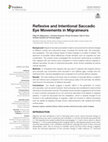
Background: Migraine has been postulated to lead to structural and functional changes of differen... more Background: Migraine has been postulated to lead to structural and functional changes of different cortical and subcortical areas, including the frontal lobe, the brainstem, and cerebellum. The (sub-)clinical impact of these changes is a matter of debate. The spectrum of possible clinical differences include domains such as cognition but also coordination. The present study investigated the oculomotor performance of patients with migraine with and without aura compared to control subjects without migraine in reflexive saccades, but also in intentional saccades, which involve cerebellar as well as cortical networks. Methods: In 18 patients with migraine with aura and 21 patients with migraine without aura saccadic eye movements were recorded in two reflexive (gap, overlap) and two intentional (anti, memory) paradigms and compared to 25 controls without migraine. Results: The main finding of the study was an increase of saccade latency in patients with and without aura compared to the control group solely in the anti-task. No deficits were found in the execution of reflexive saccades. Conclusions: Our results suggest a specific deficit in the generation of correct anti-saccades, such as vector inversion. Such processes are considered to need cortical networks to be executed correctly. The parietal cortex has been suggested to be involved in vector inversion processes but is not commonly described to be altered in migraine patients. It could be discussed that the cerebellum, which is recently thought to be involved in the pathophysiology of migraine, might be involved in distinct processes such as spatial re-mapping through known interconnections with parietal and frontal cortical areas.
Journal of Neurology, Oct 21, 2012
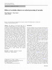
The objective of the present study was to investigate cerebellar influences on cortical component... more The objective of the present study was to investigate cerebellar influences on cortical components of saccadic eye movement programming in human subjects. In 24 patients with a localized cerebellar lesion, saccadic eye movements were recorded in different reflexive (step, gap, overlap) and intentional (anti, memory, short memory sequences) tasks and compared to 23 healthy controls. The cerebellar lesions led to impairments in different saccade parameters. Cerebellar patients tended to show hypermetria and increased latencies compared to the control group. In particular, they executed significantly more erroneous saccades specifically in the memory task (suppression errors) but not in the anti task (pro-saccade errors). Moreover, while reproducing short sequences of saccades from memory, patients with cerebellar infarcts made more errors with regard to the sequence order than controls. The influence of cerebellar hemispheric lesions on the saccade latency, the task-specific lesion effects on the frequency of suppression errors, and the effects on the number of order errors suggest that the cerebellum is involved in cortical processes such as target selection and sequence reproduction.
Pain, Jul 1, 2014
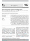
Animal studies have suggested that the cerebellum, in addition to its motor functions, also has a... more Animal studies have suggested that the cerebellum, in addition to its motor functions, also has a role in pain processing and modulation, possibly because of its extensive connections with the prefrontal cortex and with brainstem regions involved in descending pain control. Consistently, human imaging studies have shown cerebellar activation in response to painful stimulation. However, it is presently not clear whether cerebellar lesions affect pain perception in humans. In the present study, we used experimental pain testing to compare acute pain perception and endogenous pain inhibition in 30 patients 1 to 11 years after cerebellar infarction and in 30 sex-and age-matched healthy control subjects. Compared to controls, patients exhibited a significantly increased pain perception in response to acute heat stimuli (44°C-48°C, average pain intensity rating for patients 3.4 ± 2.8 and for controls 1.5 ± 1.7 [on a numeric rating scale of 0-10], P < .01) and to repeated 256 mN pinprick stimuli (1.3 ± 1.9 vs 0.6 ± 1.0 [0-10], P < .05). Heat hyperalgesia in patients was more pronounced on the body side ipsilateral to the infarction. In addition, patients showed reduced offset analgesia (change in pain intensity rating: 0.0% ± 15.8% vs À16.9% ± 36.3%, P < .05) and reduced placebo analgesia (change in pain intensity rating: À1.0 ± 1.1 vs À1.8 ± 1.3 [0-10], P < .05) compared to controls. In contrast, heat and pressure pain thresholds were not significantly different between groups. These results show that, after cerebellar infarction, patients perceive heat and repeated mechanical stimuli as more painful than do healthy control subjects and have deficient activation of endogenous pain inhibitory mechanisms (offset and placebo analgesia). This suggests that the cerebellum has a previously underestimated role in human pain perception and modulation.
Neuropsychologia, Nov 1, 2014
Decades of research have implicated both cortical and subcortical areas, such as the cerebellum, ... more Decades of research have implicated both cortical and subcortical areas, such as the cerebellum, as playing an important role in motor learning, and even more recently, in predicting the sensory consequences of movement. Still, it is unknown whether the cerebellum also plays a role in recalibrating sensory estimates of hand position following motor learning. To test this, we measured proprioceptive estimates of static hand position in 19 cerebellar patients with local ischemic lesions and 19 healthy controls, both before and after reach training with altered visual feedback of the hand. This altered visual feedback, (301 cursor-rotation) was gradually introduced in order to facilitate reach adaptation in the patient group. We included two different types of training (in separate experiments): the typical visuomotor rotation training where participants had full volition of their hand movements when reaching with the cursor, and sensory exposure training where the direction of participants' hand movements were constrained and gradually deviated from the cursor motion (Cressman, E. K., Henriques, D. Y., 2010. Reach adaptation and proprioceptive recalibration following exposure to misaligned sensory input. J. Neurophysiol., vol. 103, pp. 1888-1895). We found that both healthy individuals and patients showed equivalent shifts in their felt hand position following both types of training. Likewise, as expected given that the cursor-rotation was introduced gradually, patients showed comparable reach aftereffects to those of controls in both types of training. The robust change in felt hand position across controls and cerebellar patients suggests that the cerebellum is not involved in proprioceptive recalibration of the hand.
Experimental Brain Research, Aug 4, 2015
neglect-like clinical symptoms of CRPS patients do not involve the allocation of visual attention.
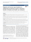
Journal of Headache and Pain, Jun 4, 2015
Background: Tension-type headache and migraine are among the most prevalent chronic disorders in ... more Background: Tension-type headache and migraine are among the most prevalent chronic disorders in children/ adolescents. Data on health care utilization for headache in this age group, however, are sparse. Methods: In 1399 grammar school students (aged 12-19 years) with headache in the last six months in Germany a) the burden of disease for headache (mean intensity, mean frequency in the last three months and PedMIDAS means), b) medical care utilization defined by proportion of students consulting a physician in the last 12 months and/or taking analgetic drugs in the last three months by headache types (migraine and tension-type headache) and by burden of disease were assessed. Results: Primary headache substantially impaired daily living activities in adolescents which was mainly related to migraine. Medical care utilization and drug use, however, was low (consulting a physician: 12.0 %, 95 %-CI = [10.3-13.8]; taking analgetic drugs: 29.9 %, 95 %-CI = [27.5-32.4])-even among students with severe headache (physician consultation: <35 %; taking analgetic drugs: <63 %). Two thirds of students with any headache and 40 % of those with migraine had neither seen a physician nor used analgetic drugs because of their headache in the preceding 12 months. Conclusions: Adolescents with headache might too rarely seek professional help for treatment of headache. Health promotion in adolescents should increase awareness for evidence-based treatment options for headache.
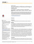
PLOS ONE, Sep 11, 2015
Objectives To assess the period prevalence and severity of dizziness and vertigo in adolescents. ... more Objectives To assess the period prevalence and severity of dizziness and vertigo in adolescents. Methods In 1661 students in 8 th-10 th grade in twelve grammar schools in Munich, Germany information on vertigo/dizziness was assessed by a questionnaire in the class room setting. Three month prevalence of dizziness/vertigo was estimated; symptoms were categorized as orthostatic dizziness, spinning vertigo, swaying vertigo or unspecified dizziness. Duration of symptoms and impact on daily life activities were assessed. Results 72.0% (95%-CI = [69.8-74.2]; N = 1196) of the students (mean age 14.5±1.1) reported to suffer from at least one episode of dizziness or vertigo in the last three months. Most adolescents ticked to have symptoms of orthostatic dizziness (52.0%, 95%-CI = [49.5-54.4], N = 863). The period prevalence for the other types of vertigo were spinning vertigo: 11.6%, 95%-CI = [10.1-13.3], N = 193; swaying vertigo: 12.2%, 95%-CI = [10.6-13.8], N = 202; and unspecified dizziness: 15.2%, 95%-CI = [13.5-17.1], N = 253. About 50% of students with spinning vertigo and swaying vertigo also report to have orthostatic dizziness. Most vertigo/dizziness types were confined to less than one minute on average. The proportion of students with any dizziness/vertigo accounting for failure attending school, leisure activities or obliging them to stay in bed were more pronounced for spinning or swaying vertigo. Conclusion Dizziness and vertigo in grammar school students appear to be as common as in adults. In face of the high period prevalence and clinical relevance of dizziness/vertigo in adolescents
Male adolescent with left-sided muscle atrophy of the hand—the rare entity of cervical flexion myelopathy (Hirayama disease)
Deutsches Arzteblatt International, Jun 11, 2021

Semiquantitative 3T Brain Magnetic Resonance Imaging for Dynamic Visualization of the Glymphatic-Lymphatic Fluid Transport System in Humans
Investigative Radiology, Apr 1, 2022
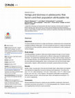
Objectives Recently, a novel clearing system for interstitial solutes of the brain was described ... more Objectives Recently, a novel clearing system for interstitial solutes of the brain was described as a perivascular pathway named the glymphatic system. Furthermore, lymphatic vessels were found in the meninges to drain interstitial fluids. It is hypothesized that interstitial solutes, such as amyloid β, are firstly processed through the brain by the glymphatic system and secondly drained out of the brain by lymphatic vessels (glymphatic-lymphatic fluid transport system [GLS]). Since then, various neurological disorders, such as Alzheimer disease, have been associated with a dysfunction of the GLS. In the current study, we aimed to establish a clinical magnetic resonance imaging (MRI) study protocol for visualizing lymphatic vessels as part of the GLS in humans. More importantly, we aimed to describe the dynamic changes of a contrast agent in these lymphatic vessels over time. Materials and Methods Twenty volunteers with an unremarkable neurological/psychiatric history were included in this 3T MRI study. Serial MRI sequence blocks were performed at 3 predefined time points (TPs): TP 1, precontrast MRI before administration of a gadolinium-based contrast agent (GBCA); TP 2, immediately post-GBCA (early ce-MRI); and TP 3, 60 minutes post-GBCA (late ce-MRI). Each MRI block contained the following sequences obtained in the same order: whole-brain 3D T1-MPRAGE, whole-brain 3D T2-FLAIR, focused 2D T2-FLAIR, and whole-brain 3D T1-SPACE. Signal intensity (SI) in compartments of the GLS adjacent to the superior sagittal sinus, gray matter (GM), white matter (WM), and cerebrospinal fluid (CSF) was calculated by manually placed regions of interest. The time course of the signal intensities was examined by generalized linear mixed models. The data were adjusted for age, cognitive function (Montreal-Cognitive-Assessment test), and sleep quality (Pittsburgh Sleep Quality Index questionnaire). Results The GLS was best visualized in the 2D T2-FLAIR and 3D T1-SPACE sequences, enabling further SI measurement. In precontrast (TP 1), the SI within the GLS was significantly higher than in CSF and significantly lower than in GM and WM. In post-GBCA, a significant increase (TP 2) and decrease (TP 3), respectively, of the GLS SI values were noted (86.3 ± 25.2% increase and subsequent decrease by 25.4 ± 9% in the 3D T1-SPACE sequence). The SI values of CSF, GM, and WM did not change significantly between the 3 TPs. Conclusions A clinical MRI study protocol was established for the visualization of lymphatic vessels as an important part of the GLS and therefore the brain’s clearing mechanism of interstitial solutes. Furthermore, dynamic changes in the GLS were described over time, possibly reflecting the clearing function of the GLS. This might constitute the basis for evaluating the GLS function in manifold neurological pathologies in the future.
PLOS ONE, Nov 13, 2017
Objectives To assess potential risk factors for vertigo and dizziness in adolescents and to evalu... more Objectives To assess potential risk factors for vertigo and dizziness in adolescents and to evaluate their variability by different vertigo types. The role of possible risk factors for vertigo and dizziness in adolescents and their population relevance needs to be addressed in order to design preventive strategies. Study design The study population consisted of 1482 school-children between the age of 12 and 19 years, who were instructed to fill out a questionnaire on different vertigo types and related potential risk factors. The questionnaire specifically asked for any vertigo, spinning vertigo, swaying vertigo, orthostatic dizziness, and unspecified dizziness. Further a wide range of potential risk factors were addressed including gender, stress, muscular pain in the neck and shoulder region, sleep duration, migraine, coffee and alcohol consumption, physical activity and smoking. Results Gender, stress, muscular pain in the neck and shoulder region, sleep duration and migraine were identified as independent risk factors following mutual adjustment: The relative risk was 1.
Jugendlicher mit linksseitigen Handmuskelatrophien – die seltene Hirayama-Flexionsmyelopathie
Clinical Neurophysiology, Oct 1, 2017
Clinical Neurophysiology, Sep 1, 2016
Scientific Reports, Oct 1, 2019
computed tomography of the heart (CCT). This technique constitutes a fast and non-invasive tool t... more computed tomography of the heart (CCT). This technique constitutes a fast and non-invasive tool that has been consistently shown to predict the risk of coronary heart disease with high accuracy 20-22. However, a relevant limitation of this approach is, that it does not directly assess non-calcified coronary plaques and coronary stenosis, thus possibly underestimating the extent of coronary heart disease. Previous studies have shown, that about one to two percent of patients with no coronary calcification at all have considerable coronary atherosclerosis due to non-calcified plaques 23,24. On the other hand, the negative predictive value of coronary artery calcifications is very high and lies between 93 and 99% 24 , which indicates an overall good estimate of coronary atherosclerosis despite its aforementioned limitations. To further investigate the common pathophysiological mechanism in migraine and coronary heart disease, as well as to clarify the association of migraine with an increased risk of cardiac events, we performed a CCT and determined the coronary artery calcium score (CACS) in migraineurs and non-migraineurs with a similar cardiovascular risk profile. Methods Patients. From July 2002 until December 2011, 1.437 Caucasian patients were included in the study. The patients underwent coronary artery calcium (CAC) scanning due to suspected coronary artery disease (CAD). Each patient signed written informed consent before CAC-scanning. The study was approved by the Data Safety Officer and the Ethics Committee of the Medical Faculty of the Ludwig-Maximilians-University Munich. All methods were performed in accordance with National Institutes of Health guidelines and regulations.
Frontiers in Neurology, Sep 28, 2022
Journal of Neurology, Jul 27, 2020
Vertigo and dizziness are amongst the most common symptoms in medicine and often have a major imp... more Vertigo and dizziness are amongst the most common symptoms in medicine and often have a major impact on activities of daily life. Although many causes of vertigo and dizziness can easily be recognized, patients often receive inappropriate and ineffective treatment. The reasons for this are various. Because vertigo/dizziness is an interdisciplinary symptom and there is a lack of standardised diagnostic tools, it is easy to lose the overview of the possible differential diagnoses. There is evidence though, that the management of patients with vertigo/dizziness can be optimized using standardized care pathways with digital support. The present study (within the framework of "PoiSe-prevention, online feedback, and interdisciplinary therapy of acute vestibular syndromes by e-health") aims to evaluate the implementation of a program with several interlocking components. The three main components are a computerized clinical decision system, a mobile application, a counselling and interdisciplinary educational program developed by the German Center for Vertigo and Balance Disorders (DSGZ). The study is a cluster-randomized controlled trial with a parallel-group design, as well as a detailed process evaluation. Clusters comprise of primary care physician practices in Bavaria, Germany. In the scope of the study the effectiveness, acceptability and efficiency of the intervention will be evaluated. It is anticipated that the intervention will improve the quality and efficiency of the management of dizzy patients. A higher diagnostic accuracy, optimized treatment, and disease progression monitoring is expected to improve patient-relevant outcomes and reduce health-care costs.
Akutes zentrales vestibuläres Syndrom
Nervenheilkunde, Feb 1, 2023
Frontiers in Neurology
BackgroundThe main clinical presentation of episodic ataxias (EAs) consists of vertigo and dizzin... more BackgroundThe main clinical presentation of episodic ataxias (EAs) consists of vertigo and dizziness attacks lasting for minutes to hours with widely varying accompanying symptoms. The differentiation of EA and episodic vertigo/dizziness syndromes in childhood and adolescence such as vestibular migraine (VM) and recurrent vertigo of childhood (RVC) can be challenging. Furthermore, only few prospective studies of children/adolescents with EA are available.ObjectiveThis study aims to characterize clinical and instrument-based findings in EA patients under 18 years of age, to delineate the clinical and therapeutic course in EA, and to present potentially new genetic mutations. Furthermore, the study aims to differentiate distinct characteristics between EA, VM, and RVC patients.MethodsWe prospectively collected clinical and instrument-based data of patients younger than 18 years, who presented at the German Center for Vertigo and Balance Disorders (DSGZ) at the LMU University Hospital ...

Uploads
Papers by Filipp Filippopulos