
Category:Proteins
biomolecule or biomolecule complex largely consisting of chains of amino acid residues  | |||||
| Upload media | |||||
| Spoken text audio | ⓘ | ||||
|---|---|---|---|---|---|
| Instance of |
| ||||
| Subclass of |
| ||||
| Part of |
| ||||
| Has part(s) | |||||
| Different from | |||||
| |||||

Subcategories
This category has the following 153 subcategories, out of 153 total.
*
1
- 14-3-3 proteins (14 F)
?
A
- Protein abundance (11 F)
- Actin-related protein 3 (1 F)
- Protein aggregation (2 F)
- Amino acid substitution (19 F)
- Ancient proteins (6 F)
- ANKRD35 (1 F)
- Argonaute proteins (7 F)
- Armadillo domain proteins (20 F)
- Aurora B (4 F)
B
- Bcl-x protein (6 F)
- Beta amyloid production (10 F)
C
- Calgranulin A (2 F)
- Calgranulin B (2 F)
- Calgranulin C (1 F)
- CCDC74A (3 F)
- Protein classes (2 F)
- Cofilin 1 (24 F)
- Protein conformation (223 F)
- Contractile proteins (169 F)
- Cortactin (13 F)
D
- Protein denaturation (14 F)
- Protein droplet (2 F)
E
- Ectodomain (8 F)
- Protein expression (9 F)
- Protein extraction (7 F)
F
- FAM63B (8 F)
- Fetal proteins (14 F)
G
- GM130 (11 F)
H
- Hemeproteins (11 F)
- Histatins (4 F)
- Homeodomain proteins (115 F)
- Protein homology detection (10 F)
I
- Protein isoforms (76 F)
L
- LIM-homeodomain proteins (8 F)
- Lithostathine (3 F)
- LOC101059915 (1 F)
M
- Mabinlin (3 F)
- Mad2 proteins (11 F)
- Matrix gla protein (6 F)
- Protein multimerization (166 F)
N
- Neoplasm proteins (37 F)
- Nerve tissue proteins (226 F)
- Neuregulin-1 (13 F)
- Nevit Dilmen Proteins (1 P, 215 F)
- Nodal protein (20 F)
- NPC1 (6 F)
- NPC1L1 (3 F)
O
- Opsonin proteins (13 F)
P
- Paxillin (27 F)
- Protein prenylation (12 F)
- Protein sequencing (5 F)
- Proteinuria (4 F)
- PrPC proteins (10 F)
Q
- Qa-SNARE proteins (9 F)
R
- R-SNARE proteins (6 F)
- Ran GTP-binding protein (6 F)
- Ras proteins (69 F)
- Resilins (7 F)
- RNA-protein interactions (20 F)
S
- Snare proteins (2 F)
- Protein stability (60 F)
- Protein subunits (49 F)
- Protein synthesis (32 F)
T
- T-box domain proteins (16 F)
- Tacrolimus binding proteins (20 F)
U
W
Z
- Zyxin (6 F)
Media in category "Proteins"
The following 200 files are in this category, out of 1,273 total.
(previous page) (next page)-
-
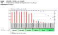
 -16 Frequency Polygon.JPG 710 × 610; 23 KB
-
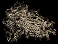
 005-CytochromecOxidase-cytox composite-1oco 1qle.tiff 1,660 × 810; 3.85 MB
-
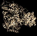
 1-s.png 1,828 × 1,392; 2.23 MB
-
 10-nsp12 apo.png 1,542 × 1,528; 1.38 MB
-
 11-nsp10.png 1,690 × 1,550; 1.11 MB
-
 12-ndp10-sinefungin.png 1,730 × 1,440; 1.21 MB
-
 12985 2016 561 Fig2 HTML.webp 778 × 579; 69 KB
-
 13-nsp15.png 1,922 × 1,632; 1.31 MB
-
 13197 2013 1042 Fig1 HTML.jpg 798 × 373; 42 KB
-
 14-orf7a.png 1,296 × 1,450; 492 KB
-
 15-orf7a.png 1,556 × 1,594; 1.14 MB
-
 18 2014 1695 Fig6 HTML.webp 387 × 348; 17 KB
-
 1a8y.jpg 1,000 × 797; 328 KB
-
 1acb.png 737 × 789; 16 KB
-
 1au1.jpg 1,000 × 797; 205 KB
-
 1B34 SmD1 8strand.JPG 1,018 × 686; 87 KB
-
 1b4r screenshot pkd DOMINIO.jpg 1,689 × 950; 113 KB
-
 1bbp.jpg 1,000 × 797; 195 KB
-
 1bbt.jpg 1,000 × 797; 265 KB
-
 1bv1.png 526 × 491; 107 KB
-
 1cd3.jpg 1,000 × 797; 302 KB
-
 1csg.jpg 1,000 × 797; 103 KB
-
 1dfn.jpg 1,000 × 797; 124 KB
-
 1dxx.jpg 1,000 × 794; 311 KB
-
 1e1w.jpg 1,000 × 794; 179 KB
-
 1efn.png 1,280 × 1,280; 794 KB
-
 1eot.jpg 1,000 × 797; 145 KB
-
 1F6R.png 1,024 × 768; 282 KB
-
 1fga.jpg 1,000 × 794; 174 KB
-
 1gof front b.png 545 × 654; 68 KB
-
 1gof front.GIF 545 × 654; 61 KB
-
 1jxu.jpg 1,000 × 794; 146 KB
-
 1K36.pdb.png 200 × 350; 28 KB
-
 1lgn.jpg 1,000 × 797; 270 KB
-
 1lj7.jpg 1,000 × 797; 306 KB
-
 1n9c.jpg 1,220 × 768; 345 KB
-
 1NEB.pdb.png 1,320 × 722; 194 KB
-
 1omf.jpg 1,000 × 794; 342 KB
-
 1pdg.jpg 1,000 × 797; 101 KB
-
 1poh.jpg 1,000 × 797; 172 KB
-
 1QNF Photolyase 190721.jpg 3,264 × 1,824; 1.31 MB
-
 1ryo.jpg 1,000 × 749; 159 KB
-
 1sbf.jpg 1,000 × 794; 298 KB
-
 1snx.png 512 × 474; 45 KB
-
 1svc NFkB DNA.png 800 × 800; 208 KB
-
 1T5R Protein Leukocidin S.png 2,048 × 1,156; 332 KB
-
 1T7P Crystal Structure.jpg 995 × 1,039; 263 KB
-
 1tap.jpg 1,000 × 797; 134 KB
-
 1tup.jpg 1,000 × 797; 279 KB
-
 1UVQ.png 1,440 × 1,080; 464 KB
-
 1www.jpg 1,000 × 797; 251 KB
-
 1z2c asr r 500.jpeg 500 × 500; 46 KB
-
 1Z90 monomer.jpg 1,011 × 760; 61 KB
-
 1Z90 oligomer.jpg 1,011 × 760; 417 KB
-
 1ztm.jpg 1,000 × 797; 201 KB
-
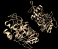
 2-S.png 2,250 × 1,572; 1.44 MB
-
2000 Antifreeze Proteins by Bill Nye for National Science Foundation.oga 1 min 3 s; 508 KB
-
 2020 08 28 N glikan z legenda.png 3,000 × 2,100; 341 KB
-
 2BC4.pdb1.png 1,436 × 759; 274 KB
-
 2cb8.jpg 1,000 × 797; 156 KB
-
 2cs4 PDB n-terminal domain chromosome 12 ORF 2.png 454 × 420; 123 KB
-
 2fjz app.png 465 × 694; 70 KB
-
 2gu0.jpg 1,000 × 794; 304 KB
-
 2GVD.gross.jpg 581 × 631; 82 KB
-
 2jsj obestatin.png 4,000 × 3,621; 1.12 MB
-
 2M9V asym r 500.jpg 500 × 500; 56 KB
-
 2mhr.jpg 1,000 × 797; 219 KB
-
 2p0a.jpg 1,000 × 794; 328 KB
-
 2QH7.png 960 × 720; 240 KB
-
 2ql1.jpg 1,000 × 794; 223 KB
-
 2r5k.jpg 1,000 × 797; 294 KB
-
 2tmv.jpg 1,000 × 797; 376 KB
-
 2w4o assembly-1.jpg 500 × 500; 56 KB
-
 2WINSurfaceRendering.png 640 × 515; 338 KB
-
 2ygd.jpg 1,000 × 797; 332 KB
-
 2yl2.png 314 × 349; 108 KB
-
 2zn9.jpg 1,000 × 794; 250 KB
-
 3' UTR folding FAM71E2.png 376 × 491; 34 KB
-
 3' UTR of SMCO3.png 600 × 467; 86 KB
-
 3-nsp3.png 1,736 × 1,336; 1.01 MB
-
 3c3v.jpg 1,000 × 797; 368 KB
-
 3D protein.jpg 825 × 1,125; 231 KB
-
 3D protein.png 400 × 400; 19 KB
-
 3D structure of zingibain enzyme.png 1,101 × 656; 407 KB
-
 3jd6.jpg 1,000 × 797; 340 KB
-
 3LFM FAT Mass and Obesity Associated (Fto) Protein.png 5,911 × 3,325; 2.94 MB
-
 3mge.jpg 1,000 × 797; 275 KB
-
 3s4eskrp1-es.jpg 676 × 514; 95 KB
-
 3trx.jpg 1,000 × 794; 201 KB
-
 3ux4.jpg 1,000 × 797; 267 KB
-
 4-6w9c.png 1,682 × 1,602; 1.11 MB
-
 4dpv.jpg 1,000 × 797; 404 KB
-
 4qyz.jpg 1,000 × 797; 316 KB
-
 4tsv bio r 250.jpg 250 × 250; 18 KB
-
 4uft.jpg 1,000 × 797; 364 KB
-
 5-nsp5.png 2,036 × 1,186; 1.36 MB
-
 5cjf assembly 1.png 500 × 500; 100 KB
-
 5cna.jpg 1,000 × 797; 274 KB
-
 5exy.jpg 1,000 × 797; 354 KB
-
 5flu.jpg 1,000 × 797; 300 KB
-
 5jzc.jpg 1,000 × 797; 302 KB
-
 5mke.jpg 1,000 × 797; 384 KB
-
 5mug.jpg 1,000 × 797; 373 KB
-
 5xqi.jpg 1,000 × 797; 330 KB
-
 5zm8.jpg 1,000 × 797; 310 KB
-
 6-nsp5.png 2,032 × 1,722; 1.57 MB
-
 6ayf.jpg 1,000 × 797; 352 KB
-
 6eho.jpg 1,000 × 797; 374 KB
-
 6wbv.jpg 1,400 × 774; 323 KB
-
 7-nsp7-8.png 1,798 × 1,080; 699 KB
-
 7ACN.jpg 775 × 650; 463 KB
-
 7ash.jpg 1,120 × 774; 464 KB
-
 7zva assembly-1.jpg 500 × 500; 60 KB
-
 8-6w4b.png 2,042 × 1,562; 1,002 KB
-
 9-nsp12.png 1,868 × 1,622; 1.64 MB
-
 9ilb.jpg 1,000 × 797; 204 KB
-
 A protein interaction network in mESCs..jpg 600 × 362; 109 KB
-

-
 A1BGgeneneighb.fcgi.png 485 × 75; 1,007 bytes
-
 A1BGprod.fcgi.png 540 × 68; 1 KB
-
 AAMDC.png 2,100 × 2,100; 1.8 MB
-
 Aars alignment.png 5,262 × 2,771; 3.87 MB
-
 Abundance over time.jpg 735 × 545; 57 KB
-
 Abundància de integrina alfa 1 en els teixits humans.jpg 2,471 × 1,517; 716 KB
-
 ActA Function.jpg 550 × 326; 60 KB
-
-
-
 AF-Q6UWT2-F1.png 426 × 213; 13 KB
-
 AF-Q9NYA4-F1 (MTMR4).png 320 × 346; 61 KB
-
 Aflibercept (Eylea®).svg 480 × 350; 77 KB
-
 Agrincartoon.png 640 × 480; 61 KB
-
 Agrincartoon2.png 430 × 325; 59 KB
-
 Ah2 nih.PNG 277 × 285; 53 KB
-
 AICAR transformylase active site.png 776 × 623; 311 KB
-
 AIMP2.png 2,800 × 2,800; 6.04 MB
-
 AimR bound to a DNA Operator in SPbeta Phages.png 3,076 × 2,403; 1.13 MB
-
 AK molmol1.jpeg 1,000 × 975; 104 KB
-
 AKAP2.png 300 × 300; 31 KB
-
 Alfa 2-antiplasmina.png 1,100 × 280; 55 KB
-
 Alphafold 3D c2orf80 protein.png 742 × 584; 104 KB
-
 Alphavbeta3 integrin expression in tumors.png 1,015 × 667; 591 KB
-
 Amidinotransferase structure diagram.JPG 484 × 582; 66 KB
-
 Amino Acid Multiplets in GOLGA8H.png 583 × 282; 55 KB
-
 Amino acids by property.jpg 2,000 × 2,000; 1.19 MB
-
 Anakinra.png 588 × 676; 18 KB
-
 Analytical.jpg 640 × 400; 12 KB
-
 AnalyticalChipExample.png 250 × 200; 7 KB
-
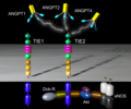 Ang-TIE-signaling.png 2,160 × 1,800; 969 KB
-
 Angiotensin-converting enzyme.jpg 483 × 335; 28 KB
-
 Anillin.jpg 1,124 × 454; 57 KB
-
 Animalian LCHN Orthologs.png 1,512 × 1,036; 525 KB
-
 ANKRD35 domains and post-transnational modifications.jpg 696 × 233; 27 KB
-
 Annotated AlphaFold Myocardin Structure.png 1,023 × 835; 217 KB
-
 Annotated conceptual translation C17orf50.pdf 1,275 × 1,650; 71 KB
-
 Annotated GrpE structure at 2.8A.jpg 4,000 × 2,250; 327 KB
-
 Annotated KIAA0090 Protein Map2.jpg 693 × 260; 24 KB
-
 Antithrombin.png 315 × 422; 30 KB
-
 AP5M1 3D Protein Structure.png 761 × 859; 159 KB
-
 APC-C substrates.jpg 650 × 531; 164 KB
-
 Apoptosi cel·lular induïda per PB1-F2.png 2,732 × 2,048; 989 KB
-
 APP cleavage mechanism.jpg 2,842 × 1,767; 1.6 MB
-
 APTX .png 2,100 × 2,100; 1.87 MB
-
 Ara h 1 structure view.tiff 604 × 600; 281 KB
-
 Ara h 1 structure.jpg 604 × 600; 363 KB
-
 ASF-SF2.png 250 × 250; 62 KB
-
 Asheetpoly.png 696 × 637; 278 KB
-
 Atatchy.png 516 × 357; 112 KB
-
 ATM 00000.png 2,100 × 2,100; 4.18 MB
-
 ATR .png 2,800 × 2,100; 4.62 MB
-
-
 B-Raf Ser-Thr Kinase Inactive Conformation.png 865 × 977; 431 KB
-
 Bacterial DIC.png 470 × 587; 240 KB
-
 Bactériorhodopsine.jpg 399 × 541; 68 KB
-
 BAR Domain on Membrane.jpg 500 × 196; 18 KB
-
 Barstar-barnase-1brs.png 708 × 504; 164 KB
-
 Baseline Sequence of 26 Repeats.png 678 × 186; 15 KB
-
 BAT5 protein human Amino Acid Sequence compared to BAT5 protein mice Amino Acid.png 2,252 × 1,256; 2.19 MB
-
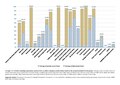
 BAT5 protein profiling in humans (h) compared to mice (m).png 1,600 × 1,262; 790 KB
-
 BAX protein 1F16.png 1,352 × 1,086; 621 KB
-
 Bcl-2 3D.jpg 400 × 376; 15 KB
-
 Bcl-2 Family.jpg 1,134 × 954; 98 KB
-
 Bcl-2 family.svg 2,601 × 2,328; 171 KB
-
 Bcl2 inhibition of Bax on mitochondrion.png 1,370 × 1,059; 84 KB
-
 Beljankovine.jpg 1,536 × 2,048; 510 KB
-
 BEND2 Diagram.png 640 × 72; 8 KB
-
 Bestrophin 1 Side edge centered.png 646 × 446; 238 KB
-
 Bestrophin-1 5T5N Xray Cystallography.png 503 × 449; 220 KB
-
 Bestrophin-1 Biolgical Assembly 1.png 519 × 524; 224 KB
-
 Beta-galactosidase (1tg7).png 500 × 500; 297 KB
-
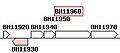 Bh11960.jpg 222 × 98; 5 KB
-
 Binding of anti-cancer drug gemcitabine-squalene to human serum albumin.png 4,302 × 4,002; 10 MB
-
 Binding of SmeT to DNA.png 603 × 539; 166 KB
-
 Bindung Fe Phosvitin V7.svg 640 × 452; 20 KB
-
 Biochemical Protein Purification Challenge.jpg 2,850 × 2,364; 1.74 MB
-
 Biological Amines.png 386 × 238; 6 KB
-
 BIOTIN.jpg 488 × 447; 29 KB
-
 Bisterpy.jpg 330 × 133; 6 KB
-
 BLAST Hemoglobin Beta.png 1,280 × 1,024; 194 KB
-
 Bloom syndrome protein.png 2,100 × 2,100; 793 KB


_Protein.png)


.png)
.svg)


_compared_to_mice_(m).png)


.png)


